Extra Foldscope Photos (2nd Post)
 May 31, 2019 • 3:22 PM UTC
 United States
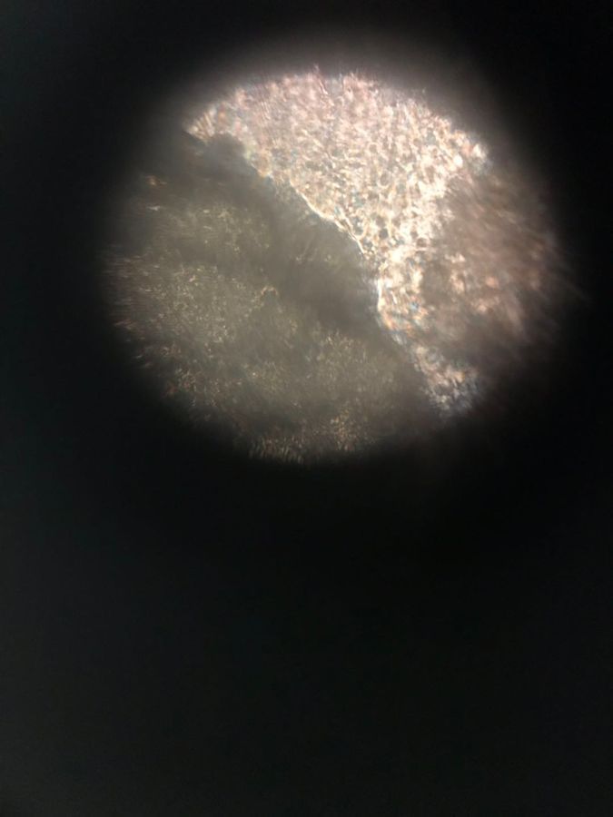
 140x Magnification
 Unknown
Shuyue Yu
Learn about the author...
4posts
0comments
3locations
View in Media Gallery
Name: Shuyue Yu
Collaborators: Happy Chang (Wenjun Chang), Catharine Long (Xiaoqi Long), Yuanzhe Xie.
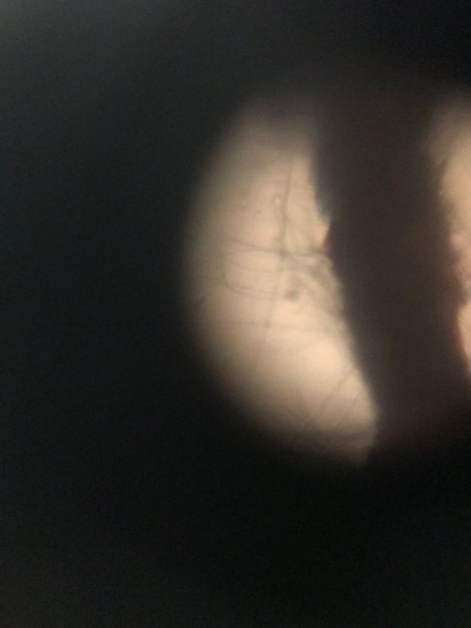
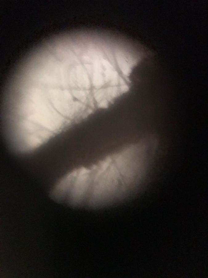
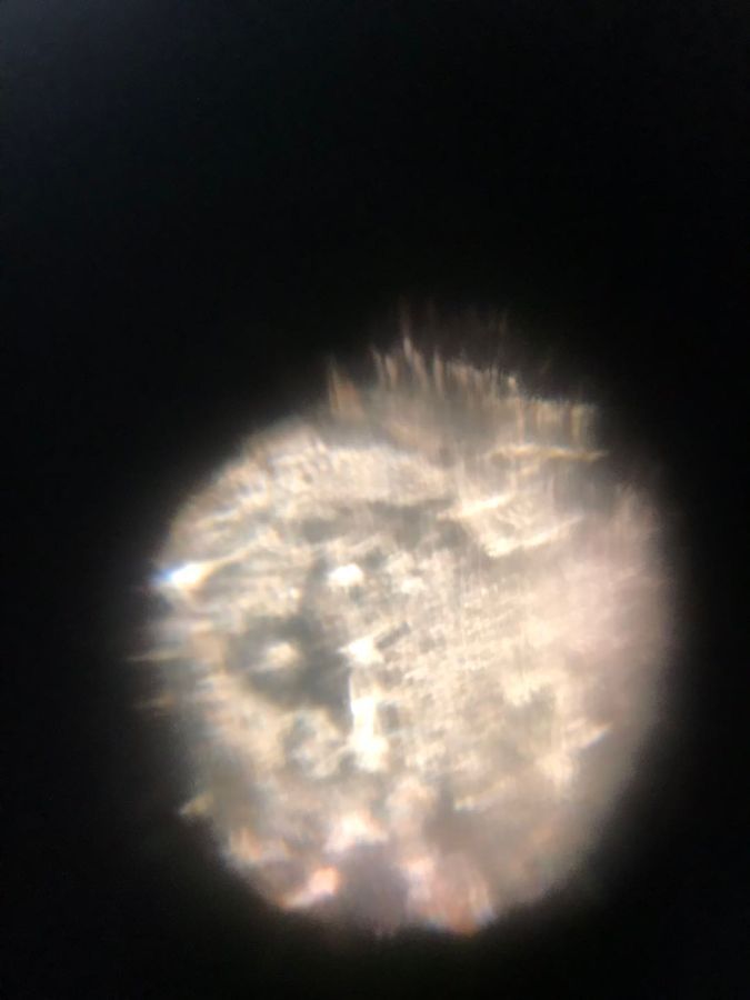
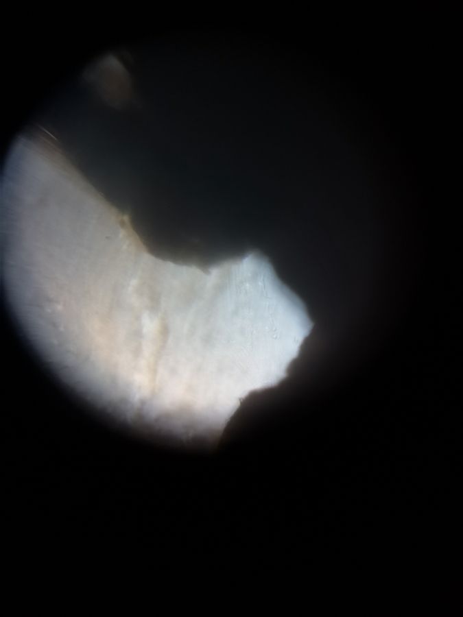
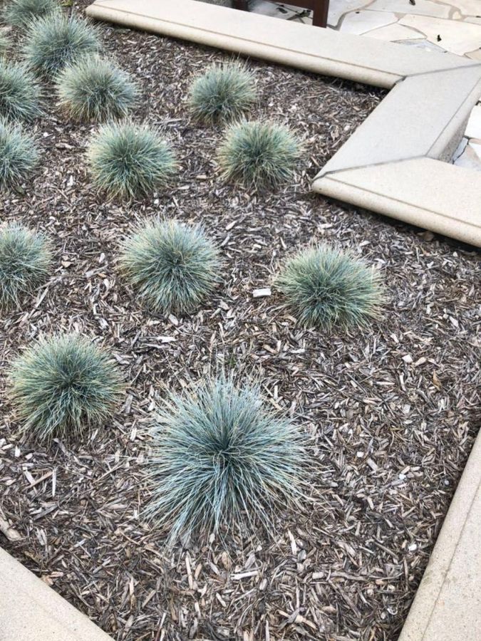
Fibers of a purple plan
On May 29th, 15:30 PM, we collected some purple plants from a bush near Annenberg at California Institute of Technology, and we got some fibers from the tip of the plant. We made the fibers of the purple plant into a sample, and the photo of it is shown below.
Collaborators: Happy Chang (Wenjun Chang), Catharine Long (Xiaoqi Long), Yuanzhe Xie.
Fibers of a purple plan
On May 29th, 15:30 PM, we collected some purple plants from a bush near Annenberg at California Institute of Technology, and we got some fibers from the tip of the plant. We made the fibers of the purple plant into a sample, and the photo of it is shown below.
View in Media Gallery
Figure 1-1. Fibers of a purple plan
View in Media Gallery
Figure 1-2. Fibers of a purple plan
View in Media Gallery
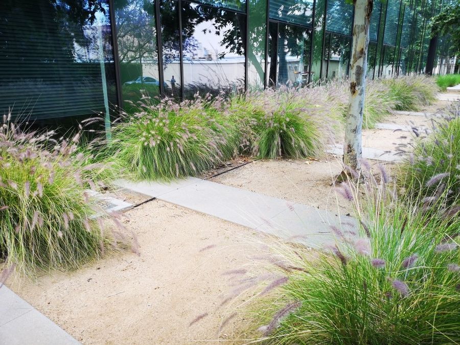
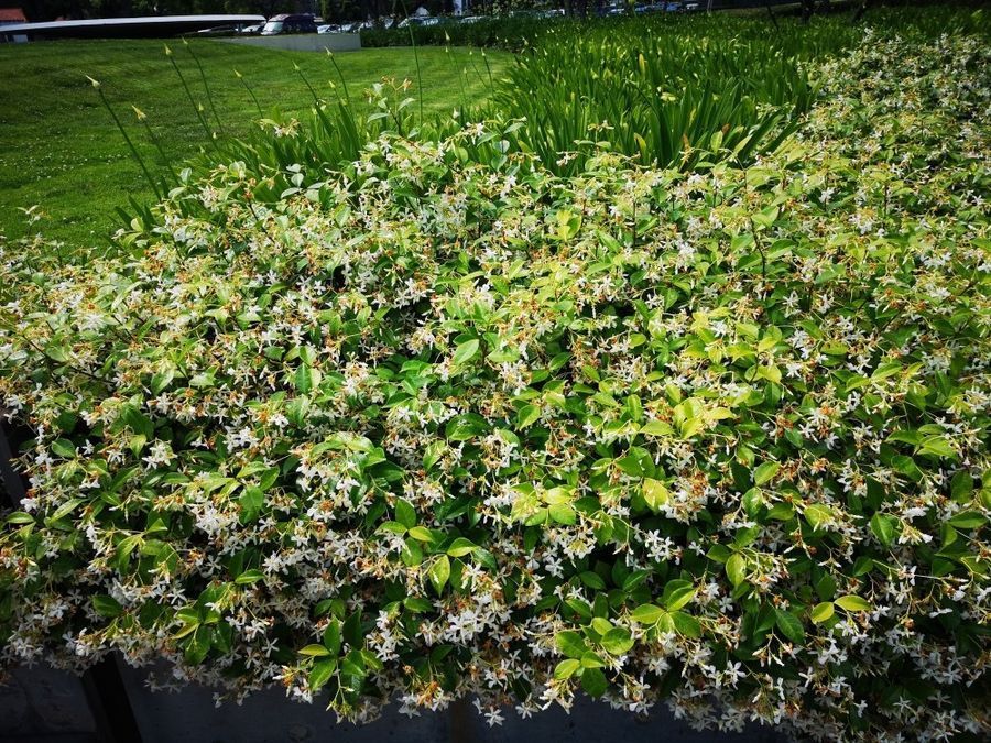
Figure 1-3. The bush near Annenburg
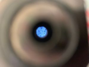
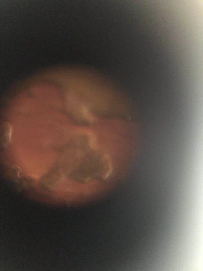
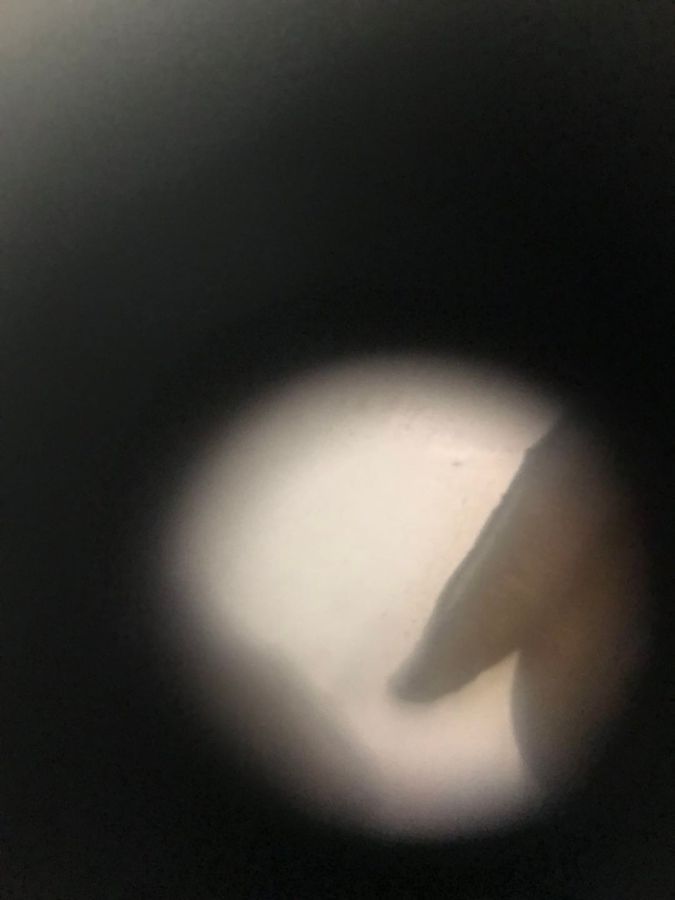
Pollen of a red flower
On May 29th, 15:32 PM, we collected some pollen from a red flower in the bush near Annenburg at California Institute of Technology. The photo of the pollen of the red flower is shown below.
Pollen of a red flower
On May 29th, 15:32 PM, we collected some pollen from a red flower in the bush near Annenburg at California Institute of Technology. The photo of the pollen of the red flower is shown below.
View in Media Gallery
Figure 2-1. Pollen of a red flower
View in Media Gallery
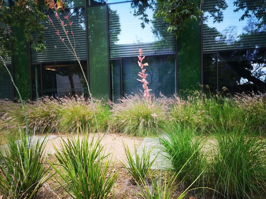
Figure 2-2. The picture of the red flower
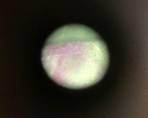
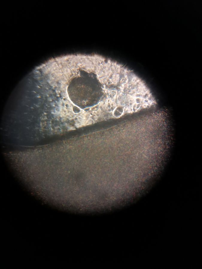
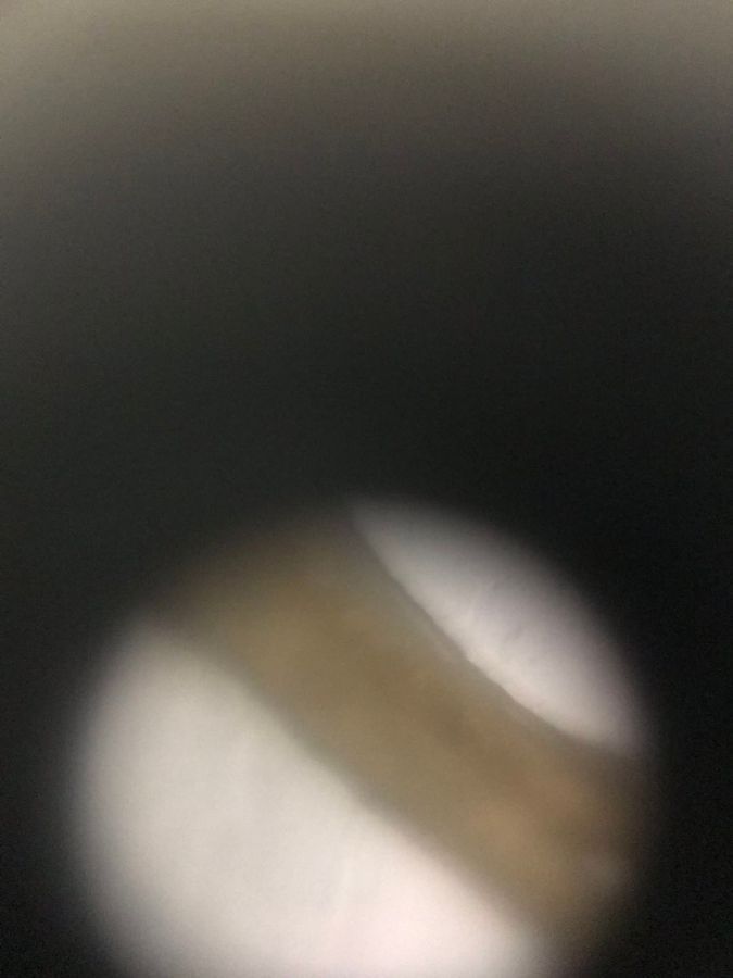
Resin of a green plant
On May 29th, 15:35 PM, we scraped some resin from the stem of a green plant in the bush near Beckman Auditorium at California Institute of Technology. The photo of the resin of the green plant is shown below.
Resin of a green plant
On May 29th, 15:35 PM, we scraped some resin from the stem of a green plant in the bush near Beckman Auditorium at California Institute of Technology. The photo of the resin of the green plant is shown below.
View in Media Gallery
Figure 3-1. Resin of a green plant
View in Media Gallery
Figure 3-2. Resin of a green plant
View in Media Gallery
Figure 3-3. The photo of the green plant
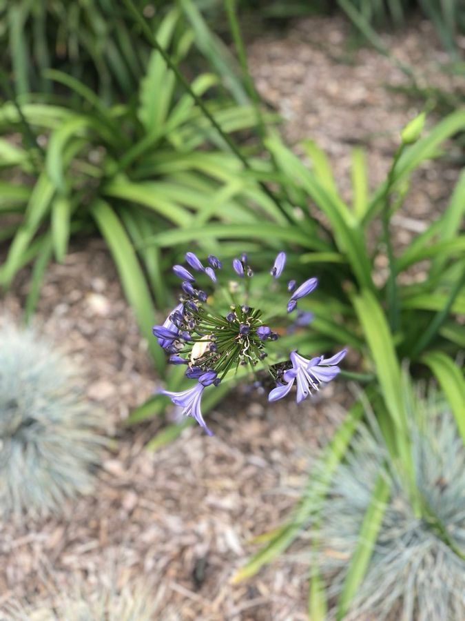
Ovule of a purple plant
On May 29th, 13:38 PM, we collected a purple flower from the garden outside Moore at Caltech. We cleaned the flower, took its ovary, and made it into a sample. The photo of the ovule of the flower is shown below.
Ovule of a purple plant
On May 29th, 13:38 PM, we collected a purple flower from the garden outside Moore at Caltech. We cleaned the flower, took its ovary, and made it into a sample. The photo of the ovule of the flower is shown below.
View in Media Gallery
Figure 4-1. Ovule of a purple flower
View in Media Gallery
Figure 4-2. The photo of the purple flower
Anther of the purple flower
Using the same flower (see Figure 4-2) that we collected on May 29th, 13:38 PM, from the garden outside Moore at California Institute of Technology, we took the anthers from the filaments and made it into a sample. The photo of the anthers of the purple flower is shown below.
View in Media Gallery
Figure 5. Anthers of the purple flower
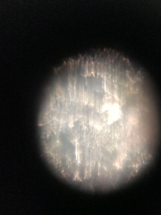
Tip of a leaf of a yellow plant
On May 29th, 13:40 PM, we took a leaf of a yellow plant at the garden outside Moore at California Institute of Technology, and we made the tip of the leaf into a sample. The photo of the tip of the leaf of the yellow plant is shown below.
Tip of a leaf of a yellow plant
On May 29th, 13:40 PM, we took a leaf of a yellow plant at the garden outside Moore at California Institute of Technology, and we made the tip of the leaf into a sample. The photo of the tip of the leaf of the yellow plant is shown below.
View in Media Gallery
Figure 6-1. Tip of the leaf of the yellow plant
View in Media Gallery
Figure 6-2. Tip of the leaf of the yellow plant
View in Media Gallery
Figure 6-3. The photo of the yellow plant
Wenjun Chang’s skin
On May 29, 13:50 PM, we got a tiny skin close to Wenjun Chang’s left index finger, and we put the skin onto the slide to make a sample. The photo of Wenjun’s skin is shown below.
Wenjun Chang’s skin
On May 29, 13:50 PM, we got a tiny skin close to Wenjun Chang’s left index finger, and we put the skin onto the slide to make a sample. The photo of Wenjun’s skin is shown below.
View in Media Gallery
Figure 7. Wenjun Chang’s skin
#caltechbi1
#caltechbi1
Sign in to commentNobody has commented yet... Share your thoughts with the author and start the discussion!
 0 Applause
 0 Comments