African fox brain (ID: 12 and 13) #BioE 301C
 May 27, 2018 • 8:04 PM UTC
 Unknown Location
 140x Magnification
 Unknown
Elle Robinson
Learn about the author...
3posts
0comments
1locations

View in Media Gallery
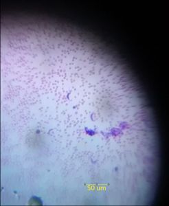
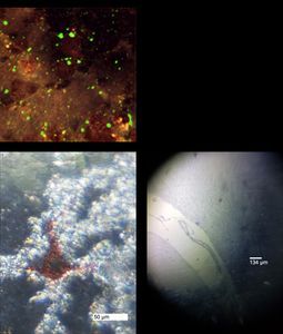
Rabies in African Fox Brain in Microscope and Foldscope. A: Immunofluorescence staining of African fox brain with rabies with 250x magnification. B: Positive test disease sample using Foldscope with a scale-bar of 50 µm. C: Negative test disease sample. This image is a rabies sample, tested positive, from an African fox brain. Associated symptoms of rabies include fever, headache, nausea, vomiting, agitation, anxiety, and confusion. Rabies, which causes inflammation in the brain, is usually fatal and is transmitted through saliva. Rabies stems from lyssavirus, a viral zoonotic neuroinvasive infectious disease transmitted by mammals that affects the CNS. Fig. 2A shows a diagnostic immunofluorescence of a fox brain with the rabies virus. Information on the staining and the original image can be found here: https://www.utmb.edu/virusimages/ . The magnification is reported to be 250x. In contrast, the scale bar is 50 µm for the positive test in Fig. 2B and 134 µm for the negative test in Fig. 2C . We used a 50 µm scale bar in Fig. 2B because it seemed to approximate the size of the red feature visible and a 145 µm scale bar in Fig. 2C because the photo was zoomed out in contrast to Fig. 2B. In Fig. 2C, we examined a larger field of view in order to see if any of the same features were visible. Features such as inflammation can be seen under the Foldscope ( https://www.cdc.gov/rabies/diagnosis/histologic.html ) in Fig. 2A. Fig. 2B shows clear inflammation similar to the pink areas in Fig. 2A. Inflamed areas were seen throughout the sample, indicating increasing spread of the rabies virus. Fig. 2B shows a sample of fox brain that does not have rabies. In this image, the sample is greatly zoomed out, but such sites indicating rabies were not visible in this sample.
Sign in to commentNobody has commented yet... Share your thoughts with the author and start the discussion!
 0 Applause
 0 Comments