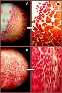
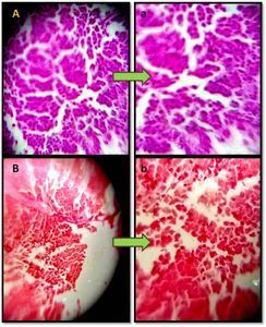
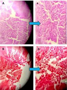
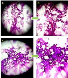
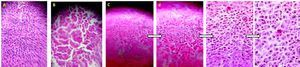
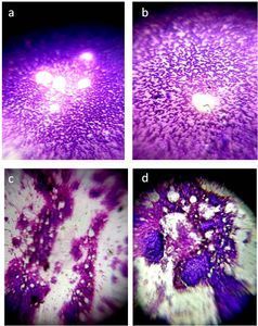
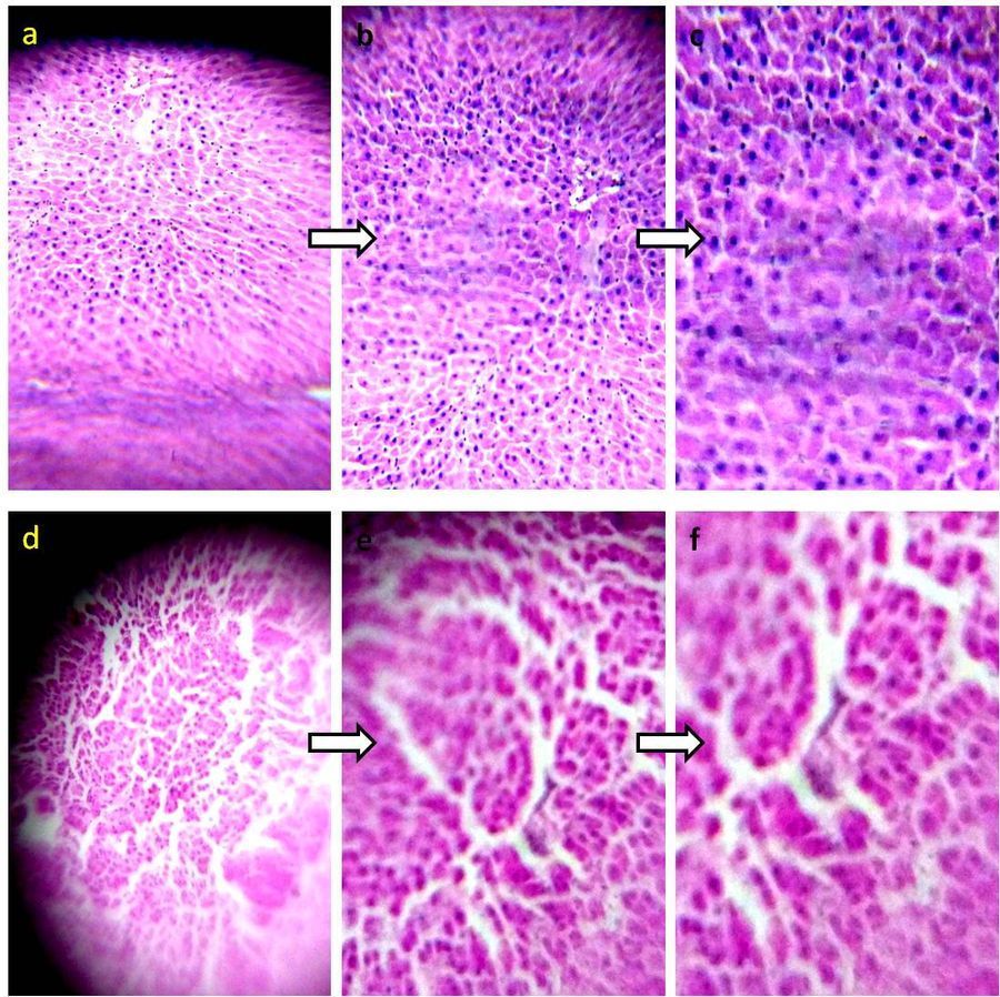
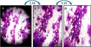
Photomicrographs of H & E stained Rat liver section –DEN induced hepatocarcinogenesis .
 Sep 04, 2018 • 3:23 AM UTC
 Unknown Location
 140x Magnification
 Microorganisms
Anindita Tripathy
Learn about the author...
8posts
0comments
1locations
View in Media Gallery
Photomicrographs of H & E stained Rat liver section –DEN induced hepatocarcinogenesis . Panel a shows untreated rat liver sections, with normal liver architecture, radiating hepatic cords surrounding the central vein. b and c images are zoom out(b- 2.5X and c-4X) images of panel a. Panel d shows DEN treated rat liver sections, with disorganized liver architecture, dilated sinusoids. e and f images are zoom out (e-2.5X and f-4X) images of panel d. Zoom out images were taken from Lenovo K6-Note android phone.
Sign in to commentNobody has commented yet... Share your thoughts with the author and start the discussion!
 0 Applause
 0 Comments