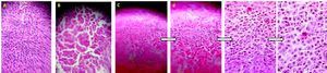
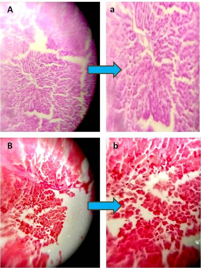
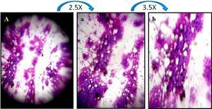
DEN induced hepatocarcinogenesis Rat liver section with Oil red-O staining.
 Sep 12, 2018 • 2:36 AM UTC
 Unknown Location
 140x Magnification
 Microorganisms
Anindita Tripathy
Learn about the author...
8posts
0comments
1locations
View in Media Gallery
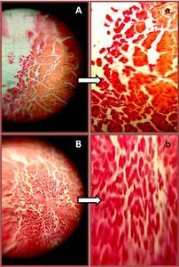
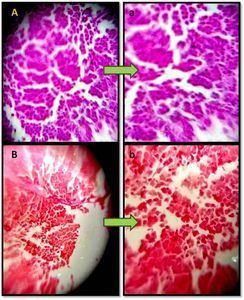
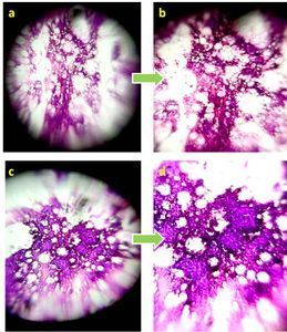
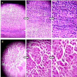
Photomicrographs of Oil red O stained Rat liver section –DEN induced hepatocarcinogenesis . Panel A shows untreated rat liver sections, with normal liver architecture. Panel B shows DEN treated rat liver sections, with Oil red-O staining. In untreated liver section Panel A it is negative staining but the staining is positive in Panel B as the lipogenic molecules are more active in DEN treated sample. a & b images are zoom out (3X) images of panel A & B respectively.
Sign in to commentNobody has commented yet... Share your thoughts with the author and start the discussion!
 0 Applause
 0 Comments