Tutorial: How to do “in-situ” live imaging of plant leaf cells without damaging a plant
 Dec 26, 2017 • 10:10 PM UTC
 Unknown Location
 140x Magnification
 Microorganisms
Manu Prakash
I am a faculty at Stanford and run the Prakash Lab at Department of Bioengineering at Stanford University. Foldscope community is at the heart of our Frugal Science movement - and I can not tell you how proud I am of this community and grassroots movement. Find our work here: http://prakashlab.stanford.edu
266posts
1192comments
42locations

Usually, the most creative (and difficult) process of live imaging is discovering how to grow/keep alive whatever you want to image under a glass slide. This is not trivial, since organisms sense local environment which has a dramatic effect on the sample.
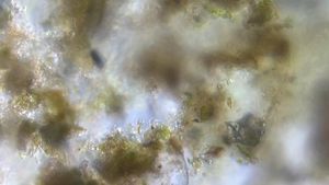
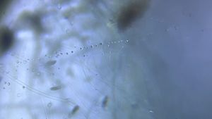
In the latest design, we coupled a new way to mount live plant samples – by magnetically coupling leaves so you can image stomatal dynamics. Here is a quick tutorial video to do the same. Depending on what you are imaging, it’s helpful to have light on maximum intensity (without diffuser). If the image is not focused; play with the new ramp.
The real thrill of this is when you can setup time lapse; go to sleep and watch a leaf grow when you wake up in the morning. Just setting it up; will update tomorrow on the same.
A post shared by Foldscope Instruments, Inc (@teamfoldscope)
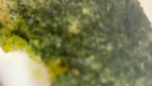
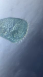
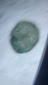
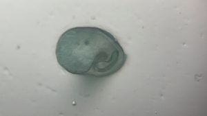
Just some quick observations. I find it remarkable how much the cellular geometry changes with every few millimeters.


Cheers
Manu
Manu


Sign in to commentNobody has commented yet... Share your thoughts with the author and start the discussion!

 0 Applause
 0 Comments