LGP26-B3 Day-3(Part 1)
 Jun 04, 2026 • 2:16 PM UTC
 India
India 140x Magnification
 Plants
Neeraj Sarmah
Greetings, This is Neeraj documenting his voyage of the microbial world.
3posts
0comments
0locations

View in Media Gallery
Greetings,
Today is Day 3(Part-1) of my voyage on the cellular safari. Today we explore a new island for treasure, called Tomato Flesh and Peel respectively.
Firstly, we prepare the Tomato Flesh, for which we get a small sample and slightly smash the tomato to get a thin layer.
Today is Day 3(Part-1) of my voyage on the cellular safari. Today we explore a new island for treasure, called Tomato Flesh and Peel respectively.
Firstly, we prepare the Tomato Flesh, for which we get a small sample and slightly smash the tomato to get a thin layer.

View in Media Gallery
We start with a 50x magnification of the Tomato Flesh. The cells are transparent and circular quite contrary to the cells of the peel.

View in Media Gallery
At 140x Magnification of the Tomato Flesh cells, we can clearly see transparent individual cells and the orange-tinted streaks

View in Media Gallery
The next is a 340x magnified image of the Tomato cell.
The red-orange rod structures suspended in the transparent matrix are chromoplasts containing crystallized lycopene. As a tomato ripens, its green chloroplasts lose their chlorophyll and turn into chromoplasts, synthesizing massive amounts of lycopene. Because these red pigments crystallize into sharp needles or rods within the membrane.
Finally, we take on the Tomato peel, we are presented with a small sample of tomato, from which we take the peel with the help of our nail to ensure a small sample. Also avoid scratching off the debris from the Tomato skin as that might get rid of all the cells that we are looking for.

View in Media Gallery
The above is the picture of the tomato peel at 50x magnification
The peel is orange-yellow in colour, with no intercellular space being seen, it looks like a tightly packed pavement. Such a dense arrangement was expected as the peel supposed to protect the inner fruit from any foreign harmful pathogen
The peel is orange-yellow in colour, with no intercellular space being seen, it looks like a tightly packed pavement. Such a dense arrangement was expected as the peel supposed to protect the inner fruit from any foreign harmful pathogen

View in Media Gallery
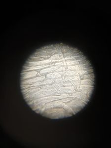
Next up we have the 140x magnified image of the tomato cells.
We can view the inter-locking mosaic quite clearly
We can view the inter-locking mosaic quite clearly

View in Media Gallery
Next up for the peel we got the 340x magnified image of the Tomato Peel.
The problems I faced today were almost none, as I had learned quite a lot in the past days regarding fine adjusting and taking thin samples.
Though I am yet to improve on taking better pictures, which I hope to get better with time.
Citations
Science Learning Hub. "Fruit Anatomy and Ripening." Science Learning Hub , 8 Nov. 2021, www.sciencelearn.org.nz/resources/2802-fruit-anatomy-and-ripening.
ThoughtCo. "Plant Cell Structures and Organelles." ThoughtCo , 12 Mar. 2023, www.thoughtco.com/plant-cell-anatomy-373620 .
Sign in to commentNobody has commented yet... Share your thoughts with the author and start the discussion!

 0 Applause
 0 Comments