LGP26-B3
 Jun 04, 2026 • 2:38 AM UTC
 India
India 140x Magnification
 Plants
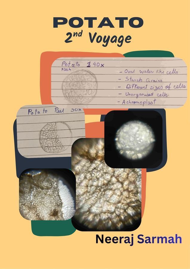
Neeraj Sarmah
Greetings, This is Neeraj documenting his voyage of the microbial world.
3posts
0comments
0locations
View in Media Gallery
This is my day 2 of the voyage into the microbial world.
Today I explored potato cells. Just like Day 1, I am ready to take on the seas, as I ride the waters to go beyond today.
Today I was presented with a small potato sample with its peel and flesh, both examined differently.
Firstly for the peel, we need to use our god-given gift, i.e our fingers to scratch up a small part of the peel from the potato and tape it nicely in place.
Then we may use our foldscope thoroughly to look at every periphery of the tissue for the best view.

View in Media Gallery
First up is the Peel of the Potato at 50x Magnification.

View in Media Gallery
Here we could see the dense cork cells all packed together and have an angular shaped irregular compartment. We can observe a brown mesh-like pattern. Also if we compare them with the flesh of the potato then, it is seen that the peel cells are noticeably smaller and more compact.
Now we move to the 140x magnification to view the cells of the potato peel via the Foldscope.
Now we move to the 140x magnification to view the cells of the potato peel via the Foldscope.

View in Media Gallery
The thick and dense; distinct boundaries between the cells are highly visible. Because these cells are empty and dead,we are primarily seeing the rugged, multi-layered cell walls. The texture appears rough and scale-like, which explains why potato skin feels coarse when we touch it.

View in Media Gallery
The above is the 340x magnified view of the Potato Peel Cell
Despite the individual cell boundaries being slightly blurred due to the thickness of the sample, we can still see and infer that there is no vacuole or nucleus in the cells, which takes us back to our textbooks where we learnt that these course peels have the function to provide structure and a protection.
Now it is time to move on further for the flesh of the potato.
First we prepare the slide, by taking a small piece of the potato and smash it slightly to not get a thick deposit, and finally remove any excess debris before taping it to the slide. Remember to not be greedy with the sample as too many cells stacked over each other would interfere with the translucent nature of the sample, hence not giving us our desired result.

View in Media Gallery
This is the first image at 50x magnification taken of the potato peel
We can see that the cells are circular and colourless, also the cell outlines are quite faint and thin as the cellulose cell wall is quite thin.
We can see that the cells are circular and colourless, also the cell outlines are quite faint and thin as the cellulose cell wall is quite thin.


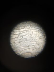
This above is the representation and picture of the 140x magnification of the Potato Flesh.
Here we can view water droplets like circular cells quite clearly, we can also see the translucent achromoplast nature of the cells, despite the cells being packed in the sample. Also we see a 3d cell.
These are starch grains, which can be verified if we stain them with iodine, which will result in a black pigment, proving our hypothesis.

View in Media Gallery
Finally, we arrive at the 340x magnification of the Potato Flesh Cell
We have magnified to individual starch granules as we see the difference in their sizes. We can also faintly see that the gap between two large granules is filled by smaller granules.
Throughout today’s voyage the following was my primary challenge-
-I initially took a lot of samples for the flesh, hence I had to re-do to get a finer thinner sample—goes to show the iterative nature of science, that I really appreciate to have experienced first hand.
Thankyou
Citations
Artschwager, Ernst. "Studies on the Potato Tuber." Journal of Agricultural Research , vol. 27, no. 11, 1924, pp. 809–835. Bhardwaj, Vinay, et al. "Potato Periderm is the First Layer of Defence against Biotic and Abiotic Stresses: a Review." European Potato Journal , vol. 64, no. 3, 2020, pp. 1–22, https://doi.org/10.1007/s11540-020-09468-8 . Cybulski, James S., et al. "Foldscope: Origami-Based Paper Microscope." PLOS ONE , vol. 9, no. 6, 2014, p. e98781, https://doi.org/10.1371/journal.pone.0098781 .
Sign in to commentNobody has commented yet... Share your thoughts with the author and start the discussion!

 0 Applause
 0 Comments