LGP26-B3
 Jun 03, 2026 • 8:50 AM UTC
 India
India 140x Magnification
 Plants
Neeraj Sarmah
Greetings, This is Neeraj documenting his voyage of the microbial world.
3posts
0comments
0locations
View in Media Gallery
Greetings,
This is Neeraj Sarmah and this is the genesis of my voyage to search for microbial treasures across the wide-vast seas of cellular scales.
Here comes the first ever image I took using my Fold Scope.
This is Neeraj Sarmah and this is the genesis of my voyage to search for microbial treasures across the wide-vast seas of cellular scales.
Here comes the first ever image I took using my Fold Scope.

View in Media Gallery
Firstly this a Fern Rhizome cell observed at 50x magnification, the organelles are clearly visible as well defined hexagonal boxes.
Further different organelles have their centres filled with the Red tints, while the variation in shade of red is due to different degrees of absorption.
At 50x we can see a wide spectrum of organelles, mostly red-pink and some artefacts of green.
This was my first time seeing a microbial life rather than a NCERT diagram from a school textbook.

View in Media Gallery
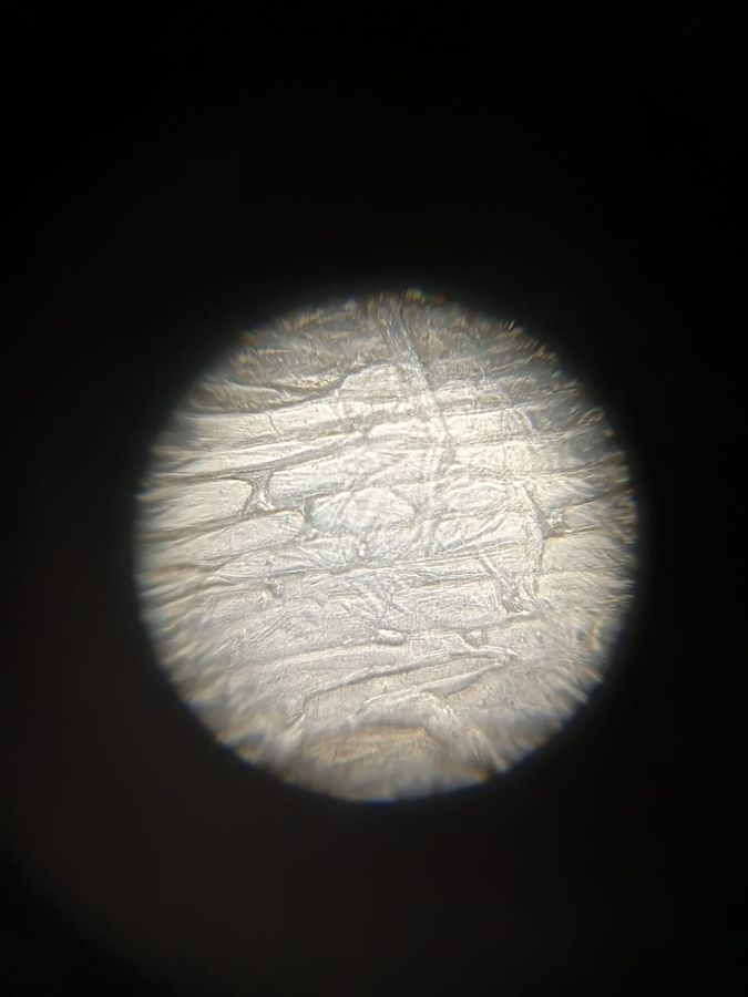
Next up we move on to the 140x magnification sample of Fern Rhizome, which gives us a more zoomed in view and a better look at the Fern Rhizome.
On a personal note, I love the 140x Fern Rhizome the most as it is the clearest photo I got.
As mentioned now, the variation of Red colour is much more clearly seen at this magnification.
On a personal note, I love the 140x Fern Rhizome the most as it is the clearest photo I got.
As mentioned now, the variation of Red colour is much more clearly seen at this magnification.

View in Media Gallery
This is the 340x Magnification of the Fern Rhizome, as seen it is much more zoomed in, though slightly on the blurry side, which I saw was a common problem for all my other peers too.
The above Fern Rhizome was already a pre-made slide on my foldscope, but now came the next step in my voyage of the cellular world, to take on the wave by preparing my own slide to observe Onion cells.
I got a small piece of Onion peel, which was thin enough for light to pass through, a pro-tip that I got while preparing the slide was to keep the samples translucent and thin for light to pass and use a cellotape to hold the onion peel firmly in place, while also avoiding any kind of moisture to avoid any future fungal growth on the foldscope.

View in Media Gallery
The above is the cool boxy looking cell of Onion at 50x magnification, where the expansive cell looks like a big mesh sheet and a brick like geometry.

View in Media Gallery
This is the 140x zoomed in picture of the onion cell, where we can see the nucleus too as small dots.

View in Media Gallery
The next is the 340x magnified image of the Onion Cell.
This concludes my DAY 1 of the Voyage on a great note that I got to learn a lot and actually go beyond my NCERT textbook, and witness a first hand experience at looking at the cells.
Though no Voyage goes in a linear fashion and hence I faced a few challenges too in getting these samples.
—Firstly, I faced initial discomfort in fine adjusting the focus to get a sharp image and a detailed look of the cells.
—Secondly, I found initial discomfort in adjusting the slide, and how thin the sample should be.
Also beyond problems I also experimented to know that the cellotape or fingerprints didn’t create any disturbance in viewing the samples.
This was all for today, a successful start to the microbial expedition.
Sign in to commentNobody has commented yet... Share your thoughts with the author and start the discussion!

 0 Applause
 0 Comments