LGP26-B3: Exploring Tomato Samples Through a Foldscope
 Jun 05, 2026 • 5:19 PM UTC
 India
India 140x Magnification
 Unknown
Ananya Dixit
Learn about the author...
6posts
0comments
0locations
_900x900.jpeg)
View in Media Gallery
Exploring Tomato Samples Through a Foldscope For this activity, I observed tomato peel and tomato flesh under 50x, 140x, and 340x magnifications using a Foldscope. It was interesting to discover how much detail is hidden inside a common fruit that we see every day.
Tomato Peel At 50x magnification , the peel appeared as a thin layer with a slightly uneven texture. The overall structure was visible, but individual cells were difficult to distinguish.
At 140x magnification , the cell boundaries became clearer, and I could observe how closely packed the cells were. The arrangement looked organized and compact.
At 340x magnification , the cells were much more distinct. The peel appeared like a connected network, showing how it acts as a protective covering for the tomato.
Tomato Flesh At 50x magnification , the flesh looked softer and more uniform than the peel. Small colored particles were visible throughout the sample.
At 140x magnification , the cells appeared larger, and some pigment-containing structures could be observed. The sample looked more complex than it did at lower magnification.
At 340x magnification , the details became much clearer. The cells and pigments were easier to see, revealing the structures that contribute to the tomato's color and texture.
One challenge I faced was preparing a thin sample because the tomato flesh was soft and watery. At higher magnifications, maintaining focus was difficult, and even slight movements caused the image to blur. After a few attempts, I was able to observe the sample clearly.
Sign in to commentNobody has commented yet... Share your thoughts with the author and start the discussion!

 0 Applause
 0 Comments
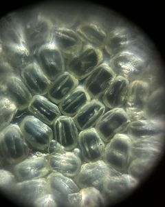

_300x300.jpeg)