LGP26-B3: Looking at an Onion Peel Through a Foldscope
 Jun 02, 2026 • 5:54 PM UTC
 India
India 140x Magnification
 Unknown
Ananya Dixit
Learn about the author...
6posts
0comments
0locations
_900x900.jpeg)
View in Media Gallery
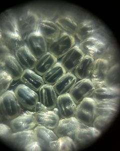
Looking at an Onion Peel Through a Foldscope During the Exploring the Microcosm session at the Lodha Genius Programme, I observed an onion peel using a Foldscope at 40×, 140×, and 340× magnifications. It was interesting to see how a common kitchen ingredient could reveal such organized structures when viewed under a microscope.
At 40× magnification, I could observe a large area of the onion peel. The cells appeared arranged in neat rows and looked similar to bricks in a wall. This magnification gave me an overall view of the tissue and showed how closely packed the cells were.
When I increased the magnification to 140×, the individual cells became much clearer. The rectangular shape of the cells was easier to identify, and the cell boundaries appeared more distinct. Since the field of view became smaller, I could see fewer cells, but I was able to notice more details within the tissue.
At 340× magnification, I was observing only a small portion of the onion peel. The cell walls appeared sharper and more detailed than before. Although I could no longer see a large number of cells at once, this magnification helped me focus on the structure of individual cells and understand how the tissue is built.
One thing I found interesting was how the same specimen looked completely different at each magnification. At lower magnification, I could appreciate the overall arrangement of the cells, while at higher magnifications, I could study the details of individual cells. This activity helped me understand why scientists use different levels of magnification depending on what they want to observe.
Overall, this observation showed me that even something as ordinary as an onion contains an organized microscopic structure. It was a reminder that there is much more to everyday objects than what we can see with our eyes alone.
Sign in to commentNobody has commented yet... Share your thoughts with the author and start the discussion!

 0 Applause
 0 Comments
_300x300.jpeg)