The Potato Peel [Day 2 LGP 26 - B2] ~ Rishi Prasad
 May 27, 2026 • 7:41 AM UTC
 India
India 340x Magnification
 Plants
Rishi Prasad
High school student
4posts
0comments
0locations
View in Media Gallery
This is a sample of a potato peel, viewed under the Foldscope 2.0. This post is aimed at sharing the observations I made during my time playing around with the sample.

View in Media Gallery
This is the peel viewed under 50x magnification. The cells visible are closely packed together and irregularly arranged. The shapes of the cells are also dissimilar to each other. Many dark spots are seen, but their exact makeup is not clear at this magnification.

View in Media Gallery
This is the same sample viewed under 140x magnification. While the cells were not very clearly visible when photographed, it was much clearer in person. Attached below is a sketch I made while viewing the sample, which shows the cells much more clearly.

View in Media Gallery
Here, the cell boundaries are much clearer, and appear rounder. The dark spots are still unclear at this magnification.


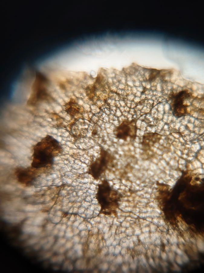
These diagrams show the same sample under 340x magnification. The cells are seen from much closer and in a lot more detail. The cell walls are much clearer. The nuclei are still not visible, but the dark spots are much clearer, appearing almost like shadows on certain cells.
I deeply enjoyed working with this sample, and I hope my diagrams and observations are of use to anyone reading this!
I deeply enjoyed working with this sample, and I hope my diagrams and observations are of use to anyone reading this!
Sign in to commentNobody has commented yet... Share your thoughts with the author and start the discussion!
![Thumbnail for Inside the Potato [Day 2 LGP 26 - B2] ~ Rishi Prasad](https://storage.googleapis.com/microcosmosdelta.appspot.com/post_media%2Fundefined%2Fundefined%2Fclonx72pfs_IMG_20260527_131518723_300x300.jpeg)
 0 Applause
 0 Comments
![Thumbnail for Inside the Onion [Day 1 LGP 26 B-2] ~ Rishi Prasad](https://storage.googleapis.com/microcosmosdelta.appspot.com/post_media%2Fundefined%2Fundefined%2F82a850rt7r_IMG_20260525_171100909_300x300.jpeg)
![Thumbnail for The Fern Rhizome [Day 1 LGP 26 - B2] ~ Rishi Prasad](https://storage.googleapis.com/microcosmosdelta.appspot.com/post_media%2Fundefined%2Fundefined%2Ft85pbksatw_IMG_20260525_161002510_300x300.jpeg)