Inside the Onion [Day 1 LGP 26 B-2] ~ Rishi Prasad
 May 26, 2026 • 5:29 PM UTC
 India
India 340x Magnification
 Plants
Rishi Prasad
High school student
4posts
0comments
0locations
View in Media Gallery
I made my first slide today! Using a thin layer of an onion that I peeled by hand and placed on the slide, I observed it from under three different magnifications. Here are my observations.

View in Media Gallery
50x magnification. Observe the thin, long rectangular cells. The cell walls' rigid structure holds this rigid structure.

View in Media Gallery
Viewed under 140x magnification. The cells are much more clearly visible now. In some, you are able to make out the outlines of the nuclei. The dark irregular shapes are the outlines of the onion's juice, caught between the onion and the slide.

View in Media Gallery
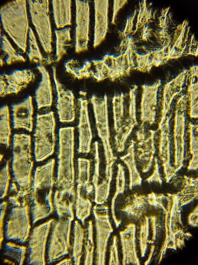
Now it is being viewed under 340x magnification. You are able to clearly make out the cell wall. The outline of the nuclei are clearly visible, and a faint outline of the sap vacuole was seen in person. The perfect, brick-like arrangement of cells looks so much more irregular now, doesn't it? That's the beauty of microscopy and biology as a whole.
I had a great time studying this sample, and I hope this post is of value to you as a fellow member of the biology community!
I had a great time studying this sample, and I hope this post is of value to you as a fellow member of the biology community!
Sign in to commentNobody has commented yet... Share your thoughts with the author and start the discussion!
![Thumbnail for Inside the Potato [Day 2 LGP 26 - B2] ~ Rishi Prasad](https://storage.googleapis.com/microcosmosdelta.appspot.com/post_media%2Fundefined%2Fundefined%2Fclonx72pfs_IMG_20260527_131518723_300x300.jpeg)
 0 Applause
 0 Comments
![Thumbnail for The Potato Peel [Day 2 LGP 26 - B2] ~ Rishi Prasad](https://storage.googleapis.com/microcosmosdelta.appspot.com/post_media%2Fundefined%2Fundefined%2Fb6x26md5yd_IMG_20260526_162224570_300x300.jpeg)
![Thumbnail for The Fern Rhizome [Day 1 LGP 26 - B2] ~ Rishi Prasad](https://storage.googleapis.com/microcosmosdelta.appspot.com/post_media%2Fundefined%2Fundefined%2Ft85pbksatw_IMG_20260525_161002510_300x300.jpeg)