LGP26-B3: Onion Peel Experiment🧅
 Jun 03, 2026 • 7:26 AM UTC
 India
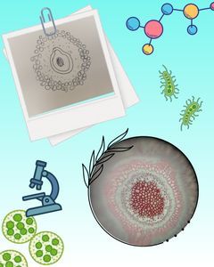
India 340x Magnification
 Plants
Aarna
Learn about the author...
5posts
0comments
0locations

View in Media Gallery
When I first placed the onion peel under the microscope, I expected to see cells. Instead, I felt like I had accidentally discovered a tiny onion mirror maze.
At 50X magnification: The peel looked shiny and reflective, almost like a microscopic mirror maze. Hundreds of cells seemed to stretch across the view in neat rows. Everything looked organized, as if the onion had hired an interior designer.
At 50X magnification: The peel looked shiny and reflective, almost like a microscopic mirror maze. Hundreds of cells seemed to stretch across the view in neat rows. Everything looked organized, as if the onion had hired an interior designer.

View in Media Gallery
At 140X magnification: Things became more interesting aaand more annoying. Getting a clear image was much harder. The cell walls became visible, and I could see that the cells were not exactly the same shape or size. They were packed together so tightly that they my clothes trying to fit in my suitcase :)

View in Media Gallery
At 340X magnification: The cells looked enormous. Unfortunately, my microscope seemed determined to test my patience. Every tiny turn of the fine adjustment knob either revealed a beautiful image or made everything blurry. Air bubbles also appeared on the slide, trying to impersonate cells, but their round shape made them easy to spot.

View in Media Gallery
In conclusion, this experiment taught me two important lessons: onion cells are surprisingly fascinating, and microscope fine-adjustment knobs have a secret mission to challenge students. :/🔬
Sign in to commentNobody has commented yet... Share your thoughts with the author and start the discussion!

 0 Applause
 0 Comments