DAY - 1 LGP 26 - B2 : FERN RHIZOME
 May 26, 2026 • 10:16 AM UTC
 India
India 340x Magnification
 Plants
Vaibhavi Sahoo
Learn about the author...
7posts
0comments
0locations
View in Media Gallery
THE ASTHETIC FERN RHIZOME : Unveiling the Hidden Geometry of Ferns
During Exploring the microcosm workshop at Ashoka University, I got an opportunity to work with Dr. Anupama and observe the Fern Rhizome under a foldscope. I honestly did not believe at first that we could use a piece of paper as a microscope but the moment I put it against my eyes, I could see the microscopic particle and that revealed something not so ordinary.
Getting to understand how the Foldscope works was an exciting experience, from adjusting the slides to the centre of the lens because apparently some people were facing a "disappearing act" while using the 340x, but jokes apart, using the fine adjustment and literally looking at the sample go from blur to clear made me so happy like a little kid seeing something fascinating for the first time. It quite literally gave my soul peace when the sample got clear. The effort? Totally worth every second.
The switching of the magnification changed my perspective a lot. The fern rhizome in particular had so many patterns and it reminded me of how something so little could be so complex beautiful and fascinating all at the same time. I'm attaching some images below of different magnifications.
50x : The bigger picture
During Exploring the microcosm workshop at Ashoka University, I got an opportunity to work with Dr. Anupama and observe the Fern Rhizome under a foldscope. I honestly did not believe at first that we could use a piece of paper as a microscope but the moment I put it against my eyes, I could see the microscopic particle and that revealed something not so ordinary.
Getting to understand how the Foldscope works was an exciting experience, from adjusting the slides to the centre of the lens because apparently some people were facing a "disappearing act" while using the 340x, but jokes apart, using the fine adjustment and literally looking at the sample go from blur to clear made me so happy like a little kid seeing something fascinating for the first time. It quite literally gave my soul peace when the sample got clear. The effort? Totally worth every second.
The switching of the magnification changed my perspective a lot. The fern rhizome in particular had so many patterns and it reminded me of how something so little could be so complex beautiful and fascinating all at the same time. I'm attaching some images below of different magnifications.
50x : The bigger picture


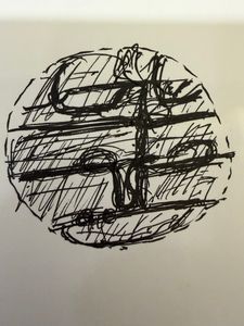
We can distinguish the major tissue systems. We can see the xylem, the phloem and the cortex.
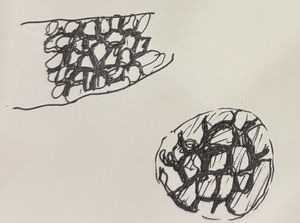
140x : Honeycomb
140x : Honeycomb

View in Media Gallery
We can literally make out the hexagonal shapes of the cells that we had earlier only read about.
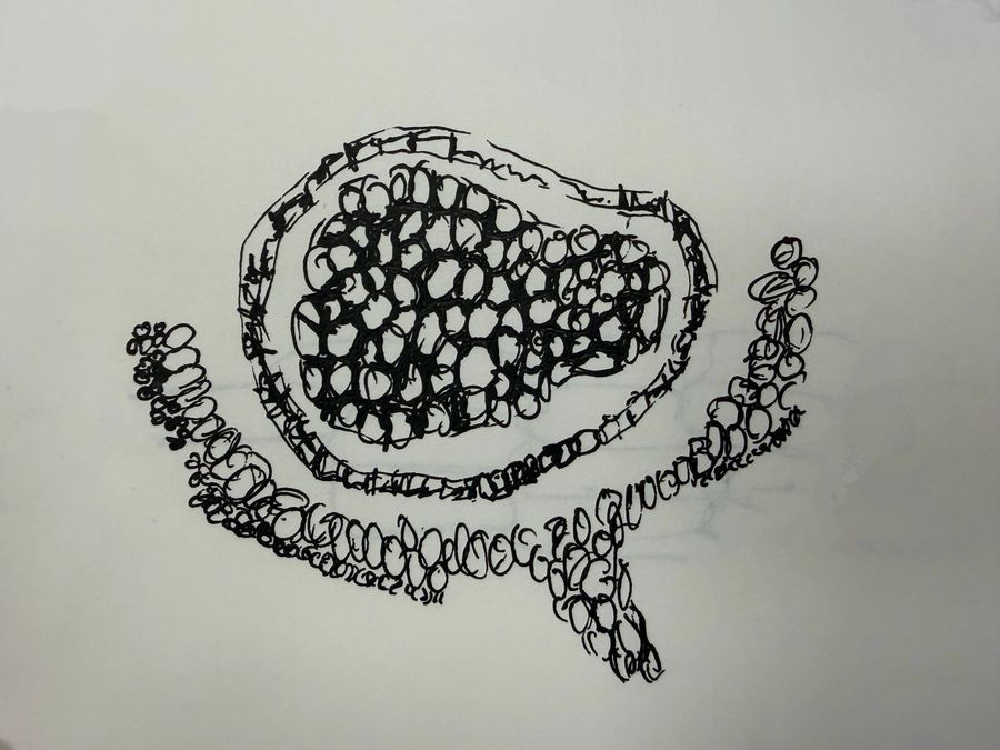
340x : The finer details
340x : The finer details


This magnification was the most fascinating to look at. The different staining made the different tissue systems stand out more.
The workshop actually changed my view about microscopic particles. Science is so much more than just memorising facts, it's about observing and exploring the real world out there.
Until my next post
See ya !!
The workshop actually changed my view about microscopic particles. Science is so much more than just memorising facts, it's about observing and exploring the real world out there.
Until my next post
See ya !!
Sign in to commentNobody has commented yet... Share your thoughts with the author and start the discussion!

 0 Applause
 0 Comments
_300x300.jpeg)