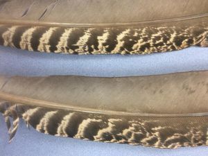
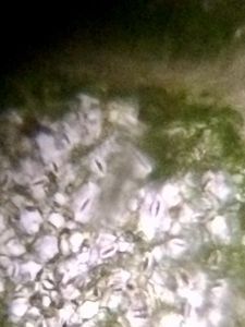
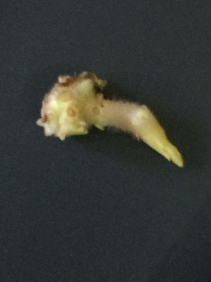
Section of potato sprout under foldscope
 Dec 20, 2018 • 10:44 AM UTC
 Unknown Location
 140x Magnification
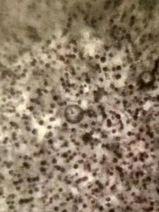
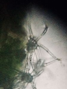
 Microorganisms
harcharan dua
Learn about the author...
17posts
0comments
1locations
View in Media Gallery
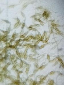
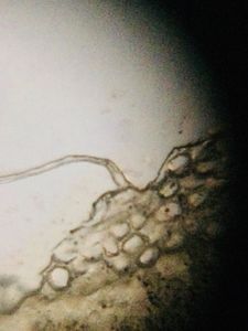
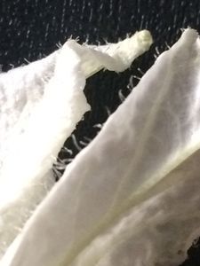
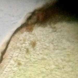
A free-hand section of a potato sprout was cut and fixed on a slide to observe the arrangement of cells anatomically under a foldscope.

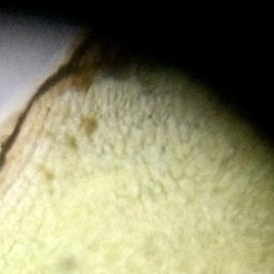
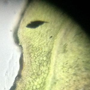
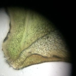
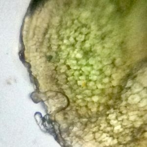
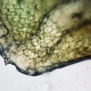
Dr. Samriti Dhawan & Dr. Jasveen Dua, GGDSD College, Chandigarh
Sign in to commentNobody has commented yet... Share your thoughts with the author and start the discussion!
 0 Applause
 0 Comments