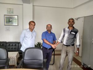
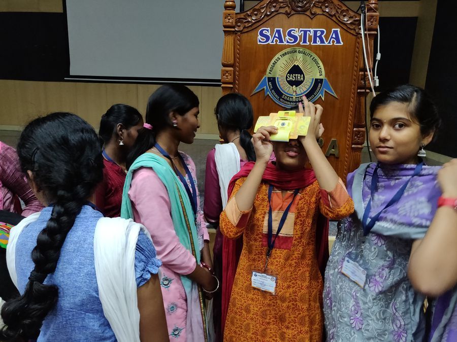
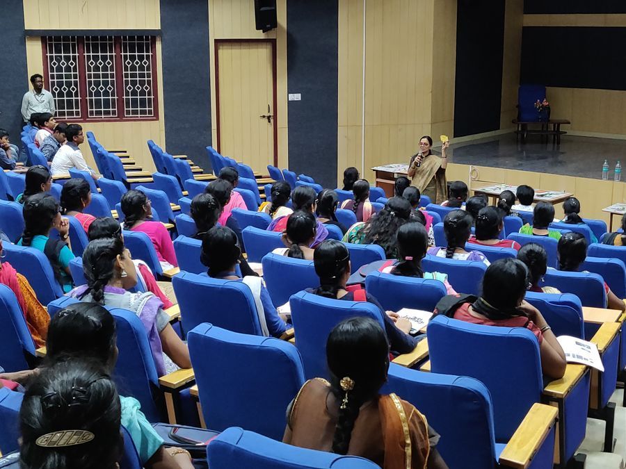
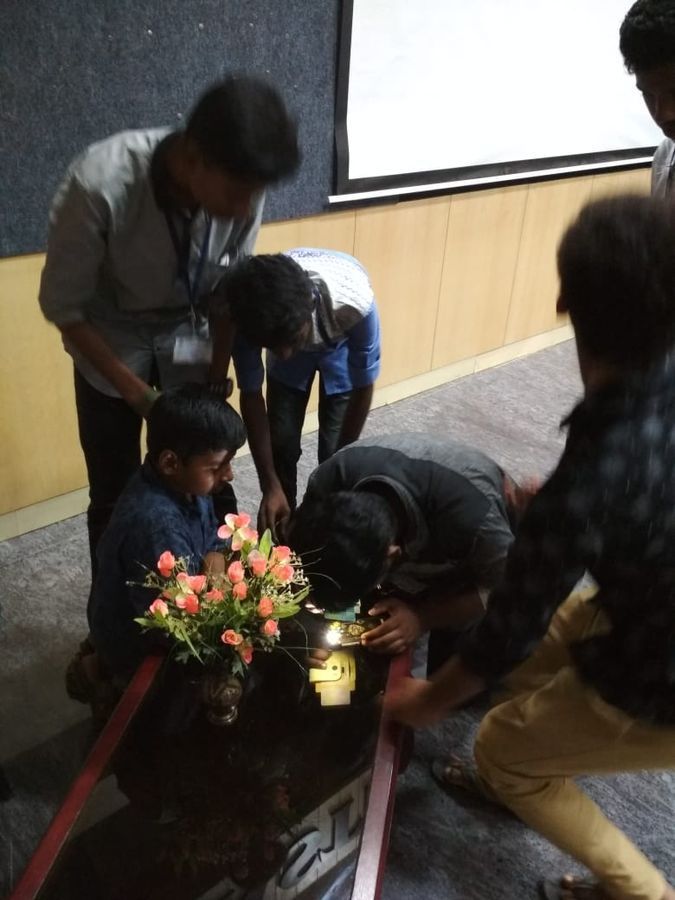
Hands-on Training using Foldscope, in DST-INSPIRE Camp at SASTRA University, on 28th Dec’18
 Dec 29, 2018 • 3:51 AM UTC
 Unknown Location
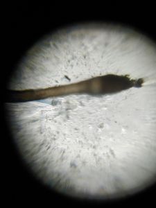
 140x Magnification
 Unknown
Raja Purushothamman
Learn about the author...
52posts
2comments
1locations

View in Media Gallery
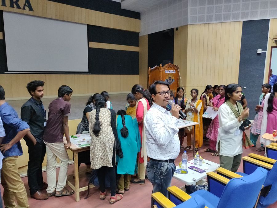
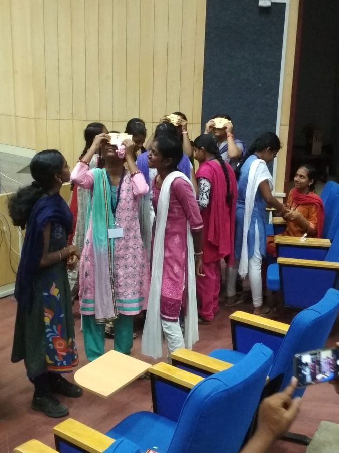
Curious listeners exploring microcosmos of biological and non-biological samples, at DST-INSPIRE camp at SASTRA University, on 28th Dec’18.
View in Media Gallery
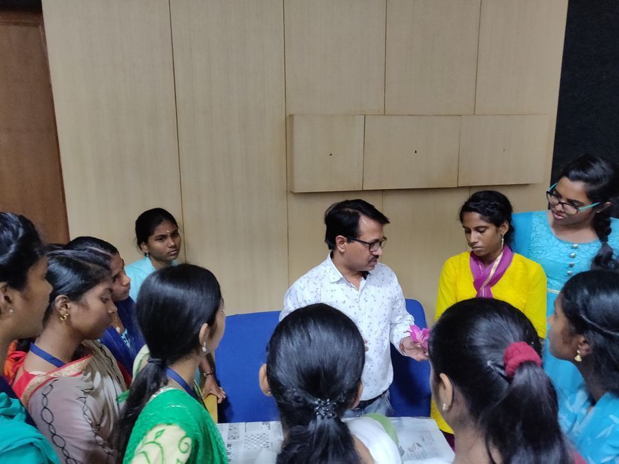
Dr. P. Raja (convener) explaining the slide insertion into the foldscope
View in Media Gallery
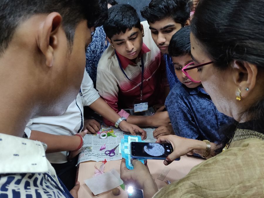
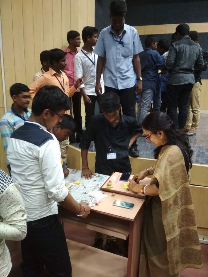
Specimen preparation instructions by Dr. N.M. Ghangaokar
View in Media Gallery
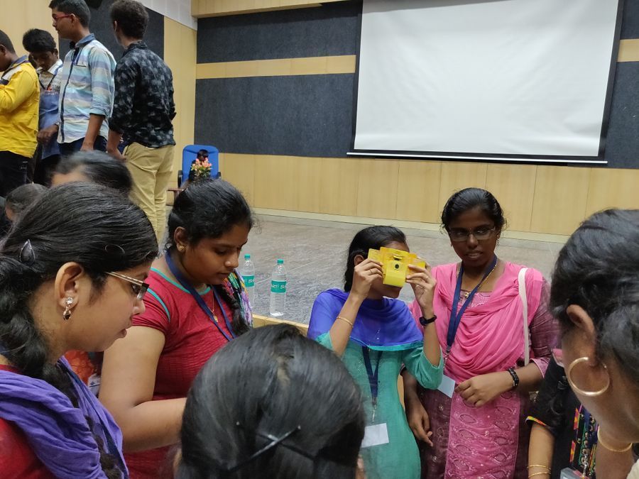
One enthusiatic student viewing the specimen slide using foldscope
View in Media Gallery
Dr. C. Deepika helping the students for viewing the specimen
View in Media Gallery
Dr. N.M. Ghangaokar explaining to students in hands on training using variuos samples
View in Media Gallery
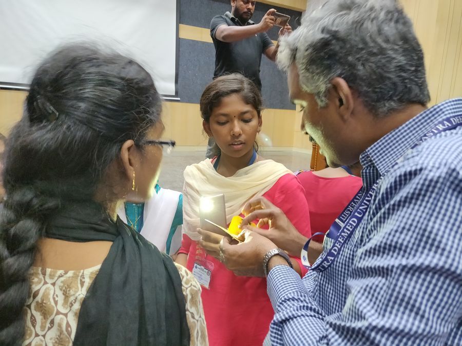
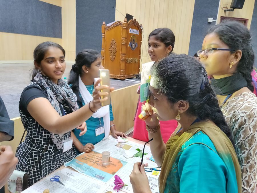
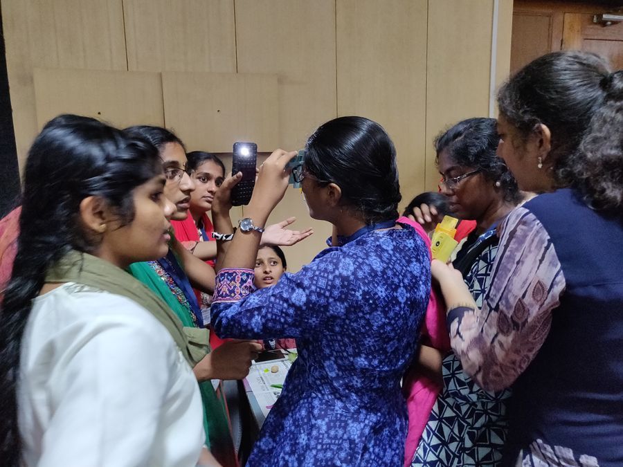
Students viewing the specimen slide using mobile torch
View in Media Gallery
Instructions to the students by Dr. Anupma
View in Media Gallery
Students viewing the bacteria in curd sample
View in Media Gallery
One student explores the specimen using Foldscope
View in Media Gallery
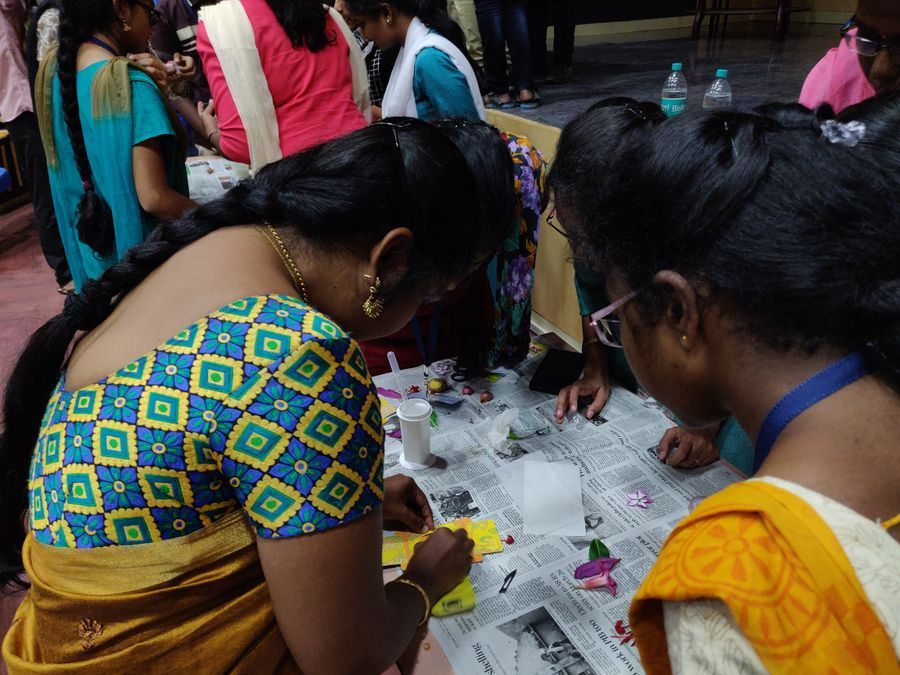
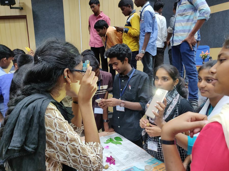
Specimen visualization using Foldscope by students
View in Media Gallery
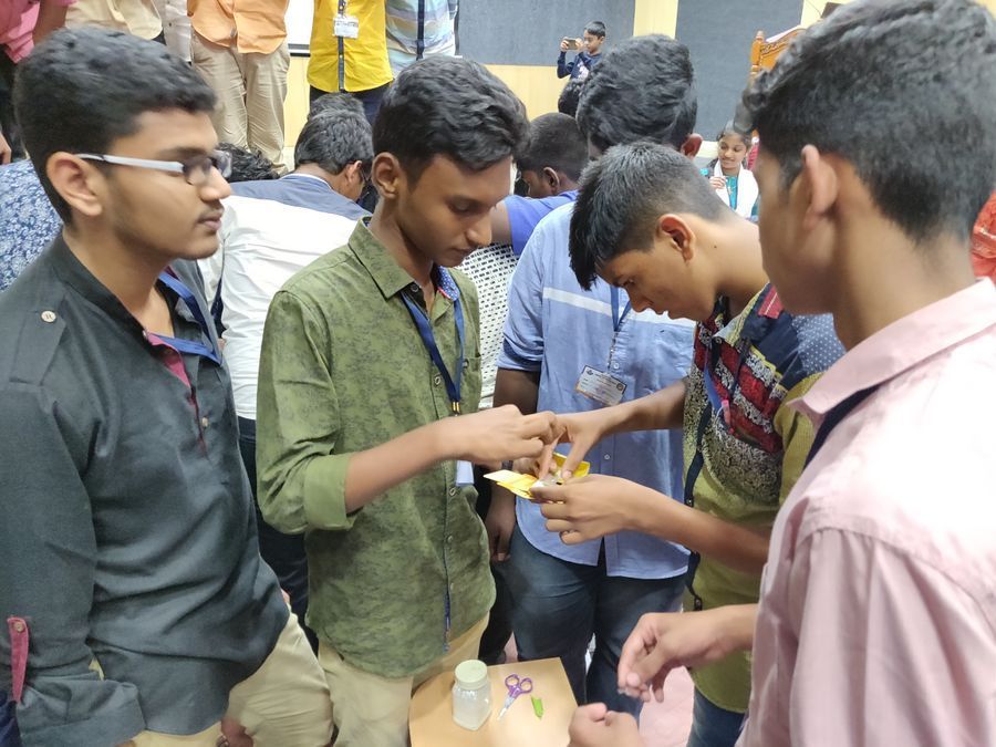
Specimen slide insertion by students
View in Media Gallery
Students enthusiastically participating in hands on training

View in Media Gallery
Exploration of specimen by students

View in Media Gallery
A curios student viewing the specimen using Foldscope
View in Media Gallery
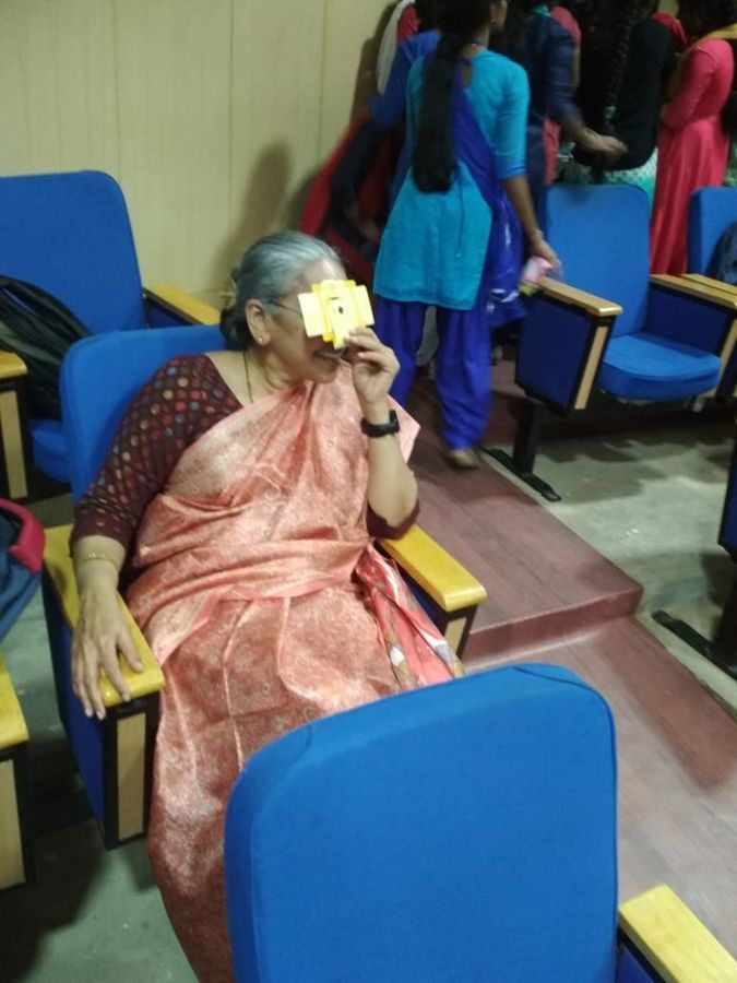
Exploring science using Foldscope by Prof. R. Vijaya from IIT-Kanpur
View in Media Gallery
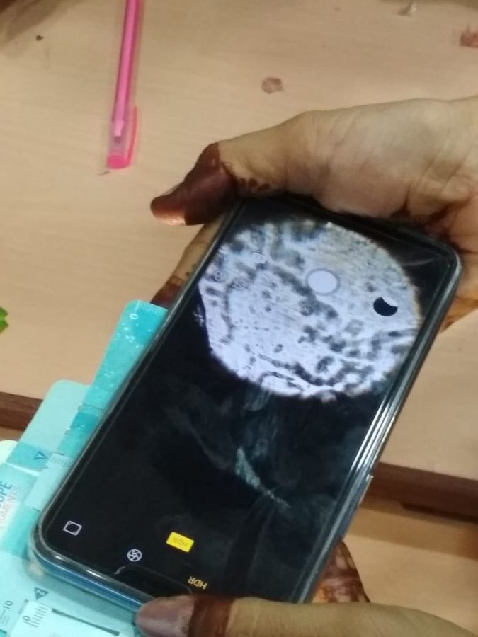
Capturing of bacteria in curd using Foldscope
View in Media Gallery
Visualization of specimen by students
View in Media Gallery
Dr. Anupma and students in hands-on training session
View in Media Gallery
Various specimens (pollen, stomata, leaf cells, curd, turbid water, etc.,) visualization by students
Sign in to commentNobody has commented yet... Share your thoughts with the author and start the discussion!
 0 Applause
 0 Comments