Ant structure under Foldscope
 Feb 24, 2019 • 10:56 PM UTC
 Unknown Location
 140x Magnification
 Unknown
Raja Purushothamman
Learn about the author...
52posts
2comments
1locations
View in Media Gallery
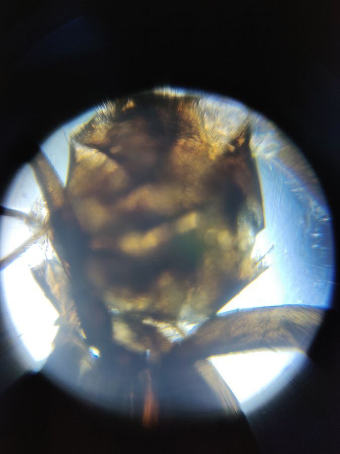
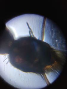
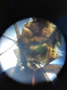
Ant body

View in Media Gallery
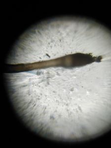
Ant leg
Ant structure under Foldscope
#Indiqfoldscopephasel.
#Indiqfoldscopephasel.
Sign in to commentNobody has commented yet... Share your thoughts with the author and start the discussion!

 0 Applause
 0 Comments