Foldscope images of plants and tissues (extra credit)
 May 30, 2019 • 11:47 PM UTC
 Unknown Location
 140x Magnification
 Unknown
Yuanzhe Xie
Learn about the author...
4posts
0comments
1locations
View in Media Gallery
This post includes 7 foldscope pictures, which are presented in the galleries below. In each gallery, the foldscope image is exhibited on the left while the right is the picture of the plant/tissue which is used to prepare the sample. These foldscope images are obtained via the collaborative efforts of a four-membered team and the team members include Wenjun Chang, Xiaoqi Long, Shuyue Yu and Yuanzhe Xie (me). The pictures displaying where the samples are obtained on the right are shared by the team members. The foldscope images are taken by each team member after the samples are prepared, which means that each team member will present unique foldscope images on the left.
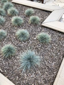
The image of the leaf of a plant (left) and the image of the location of the plant used to prepare the sample (right). The foldscope image shows a gray shadow of the leaf tip. The sample was obtained outside the Moore lab around 4 PM on May 29th. The sample was prepared by taking a small part of the leaf on squeezed by the covering glass slide.
The image of the leaf of a plant (left) and the image of the location of the plant used to prepare the sample (right). The foldscope image shows a gray shadow of the leaf tip. The sample was obtained outside the Moore lab around 4 PM on May 29th. The sample was prepared by taking a small part of the leaf on squeezed by the covering glass slide.
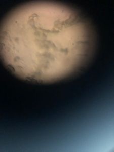
2. The image of the red flower’s pollen (left) and the image of the location of the red flower used to prepare the sample (right). The foldscope image shows small particles which correspond to pollen particles. The sample was obtained outside the Annenberg building around 3 PM on May 29th. The sample was prepared by grinding the pollen and anther in advance and covered them with the covering slide.
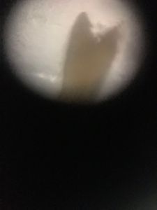
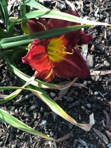
smacap_Bright 3. The anther of the purple flower (left) and the location of the plant used to prepare the sample (right). The foldscope image shows a dark outline of the anther. The sample was obtained outside the Moore lab around 4 PM on May 29th. The sample was prepared by crushing anther by the covering glass slide.
View in Media Gallery
rhdr
View in Media Gallery
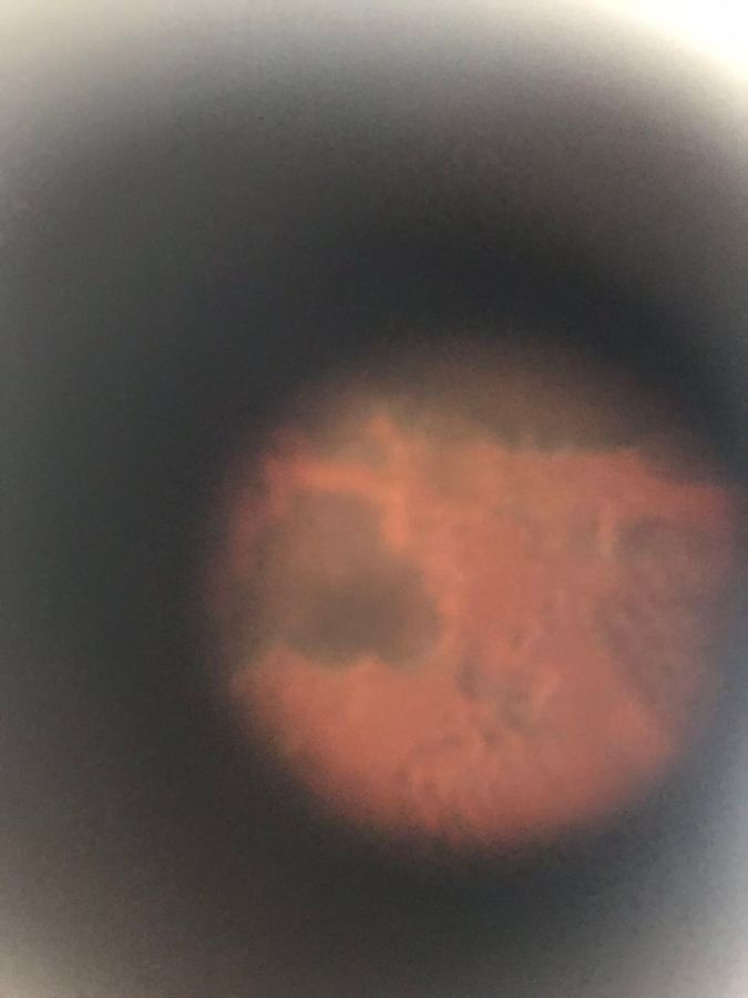
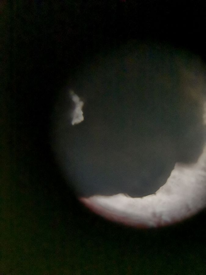
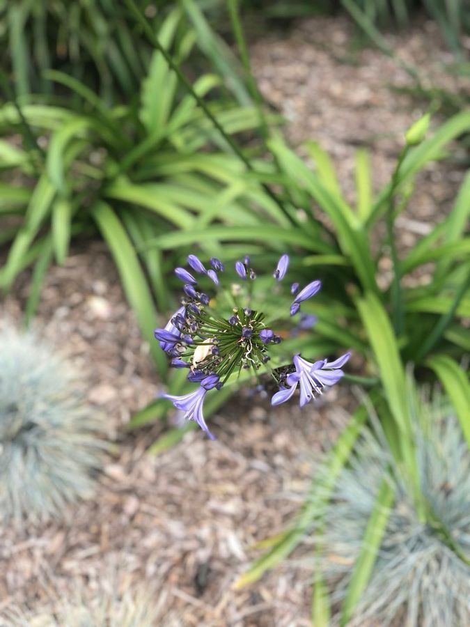
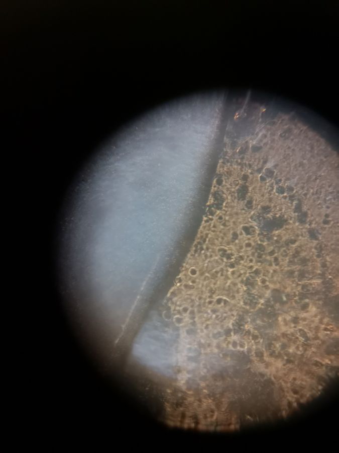
4. The ovule of the purple flower (left) and the location of the plant used to prepare the sample (right). The foldscope image shows a yellow porous structure of the ovule. The sample was obtained outside the Moore lab around 4 PM on May 29th. The sample was prepared by taking the ovule out of the flower ovary.
View in Media Gallery
mde

View in Media Gallery
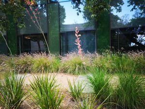
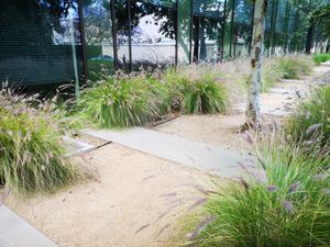
5. The filament of the bush (left) and the location of the plant used to prepare the sample (right). The foldscope image shows a dark filamentous structure of the fiber. The sample was obtained outside the Moore lab around 4 PM on May 29th. The sample was prepared by taking a small part of the grass fiber.

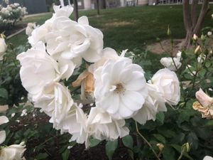
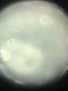
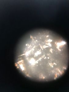
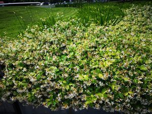
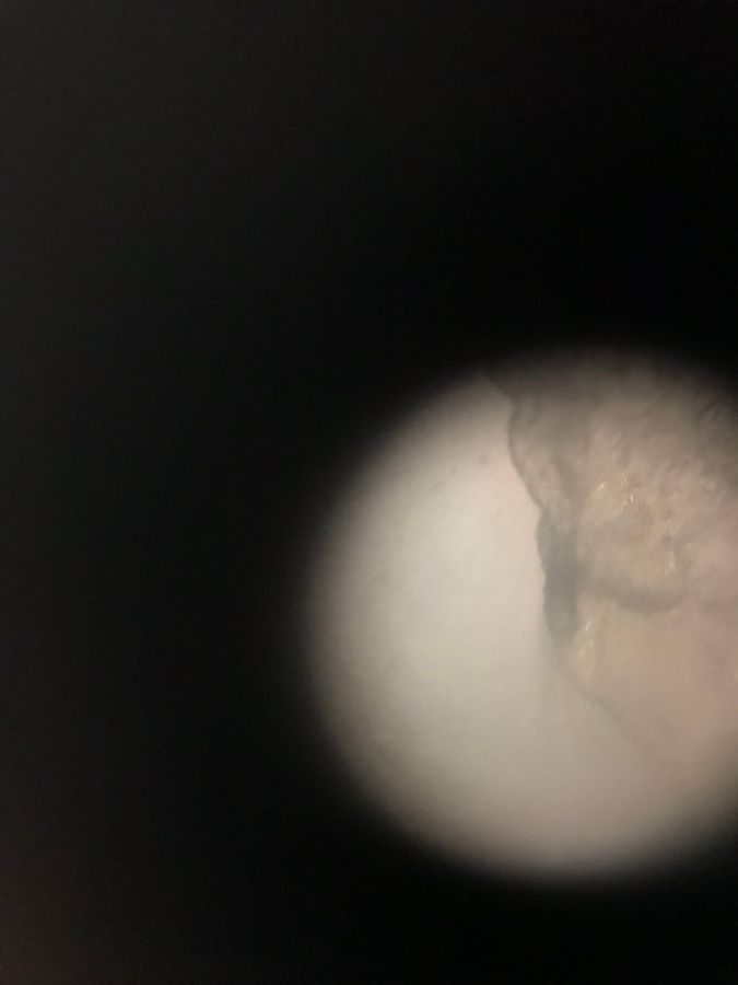
smacap_Bright 6. The resin of the white flower (left) and the location of the flower used to prepare the sample (right). The foldscope image shows a shining silver liquid. The sample was obtained around the Beckman Institute at 3:30 PM on May 29th. The sample was prepared by cutting the stem of the flower and collecting the white liquid.
smacap_Bright 7. The image of the human skin. The skin was contributed by one team member Wenjun Chang. It was ripped from his hand and covered by the covering slide. The foldscope image shows a white folded structure. The sample was obtained at 4:30 PM on May 29th in the Bechtel room 272.
View in Media Gallery
#caltechbi1
Sign in to commentNobody has commented yet... Share your thoughts with the author and start the discussion!
 0 Applause
 0 Comments