Traveling kinks – imaging bacteria under a foldscope
 Apr 04, 2017 • 11:23 PM UTC
 Unknown Location
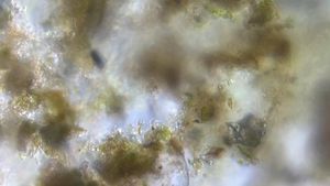
 140x Magnification
 Microorganisms
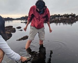
Manu Prakash
I am a faculty at Stanford and run the Prakash Lab at Department of Bioengineering at Stanford University. Foldscope community is at the heart of our Frugal Science movement - and I can not tell you how proud I am of this community and grassroots movement. Find our work here: http://prakashlab.stanford.edu
266posts
1198comments
42locations
View in Media Gallery
A million surprises are hidden right under our nose (or foot); all we often need to do is just look. And follow our nose (I mean intuition) and answers fall out of the sky. This is an optimists view of science. Let’s make good and careful observations..
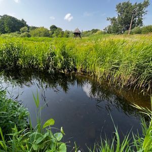
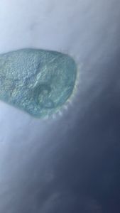
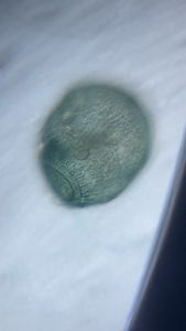
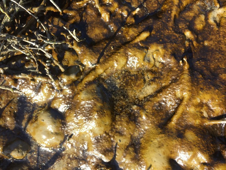
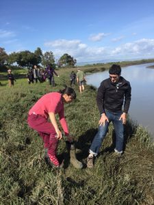
At the same swamp (Duck pond swamp, Palo Alto); just like everyone else – I also collected water samples. It’s the most unusual place; and until date – it’s the samples collected here that I have seen highest diversity of ciliates in the same sample. I will talk in details about this in a coming post; while I figure out a way to measure diversity in collected samples. In the mean time – I want to share how to image bacteria; an unusual one using a foldscope.
This tutorial assumes you have mastered the phase contrast, dark field and focus locking tricks. For revision; please see prior tutorial here:
Focuslocking, field of view locking and using ambient light (table lamp) as illumination
I took samples collected from this swamp and imaged them both in the parking lot and the same night after collecting. Yes; we did get stuck in the swamp more than once; but that was the whole point anyway. The entire group consisted of instructors, TAs of intro to biology course (coming up) at Stanford and a few dear friends from prakash lab.


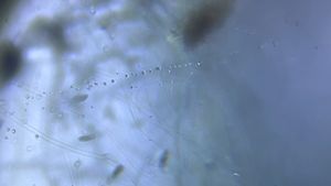
I will post all my data in an organized manner; but here is one observation – video of a swimming bacteria that should surprise you in more than one way.
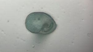
Initially you see a blank field of view; but look closely and a little helical thin rod is wiggling along the field of view. Looking at it carefully; it looks like a helical (very long) bacteria. The reason I can see this is simply because of “oblique angle illumination trick” I talked in my previous post. I often term this pseudo- phase contrast. The bacteria is clearly visible.
I think this is a “spiroplasma” – a helical Bacteria. They must come in a variety of size (thickness and length); sinc ethe current sample is almost 50um long and say 1um thick. With this oblique angle illumination; I can’t say what the smallest feature I should be able to see.
Now watch the video again; paying close attention to how could this object actually move. Watch closely and you will see “kinks” traveling fwd and backwards. It’s a subtle effect and can be seen by watching the video again and again.
Since no external appendages are visible; it should be “bending” your mind how this bacteria is actually traveling forward. But it does.. and that’s when I noticed “kinks” traveling forward and backward. When the kink travels backwards – the bacteria moves forward. Watch it slowed down and you will notice some very sharp kinks with “chirality or handedness” of the bacterial helix is reversed.
Doing some digging; I found a beautiful paper from J. Shaveitz, D. Fletcher and colleagues. Here it is attached for a future reference – I will come back to this post to show how the kinks produce active thrust forward.
Cell, 23 September 2005, Vol.122(6):941–945
Spiroplasma Swim by a Processive Change in Body Helicity
Joshua W. Shaevitz 1,,Joanna Y. Lee 2Daniel A. Fletcher 3
http://www.sciencedirect.com/science/article/pii/S0092867405006951
This is same Shaevitz from Princeton fame and Fletcher from Berkeley fame. So I am doubly excited to find this; sine I have spent short time with both of these incredible scientists.
Imaging bacteria under foldscope is a lot of fun. This was a set case since these filaments appear to be very long (as yet I can not confirm they are bacteria – my closest guess is spiroplasma; more from the motility model); but none the less very small things can be brought to light with a foldscope and a little bit of patience.
Keep exploring..
Manu
37.4608928 -122.1150112
Sign in to commentNobody has commented yet... Share your thoughts with the author and start the discussion!
 0 Applause
 0 Comments