First session with Foldscope
 May 20, 2018 • 11:54 PM UTC
 Unknown Location
 140x Magnification
 Microorganisms
Samriti Dhawan
Learn about the author...
22posts
9comments
1locations
View in Media Gallery
Reposting my first post as initially I have attached a MS word file.
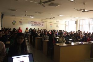
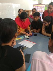
It was a great honor to be selected by Foldscope team for use of Foldscopes as a teaching and educational tool. The orientation workshop at ICGEB, New Delhi on 16 th April, 2018 gave us an opportunity to interact with our N-E twining parteners. Learning the assembly of Foldscopes from Prof. Jim and Dr. Shailja’s team at ICGEB marked the beginning of the project.
My 1 st hand experience with the foldscope
I am excited to share the results of our 1 st attempt of using Foldscope along with my Co-PI Dr. Jasveen Dua
After the orientation at ICGEB, New Delhi on assembly and usage of Foldscope, the few initial days at Chandigarh passed completing the documentation and other formalities to start working with the Foldscopes.
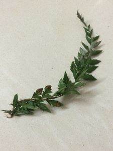
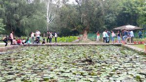
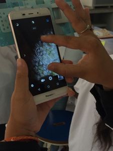
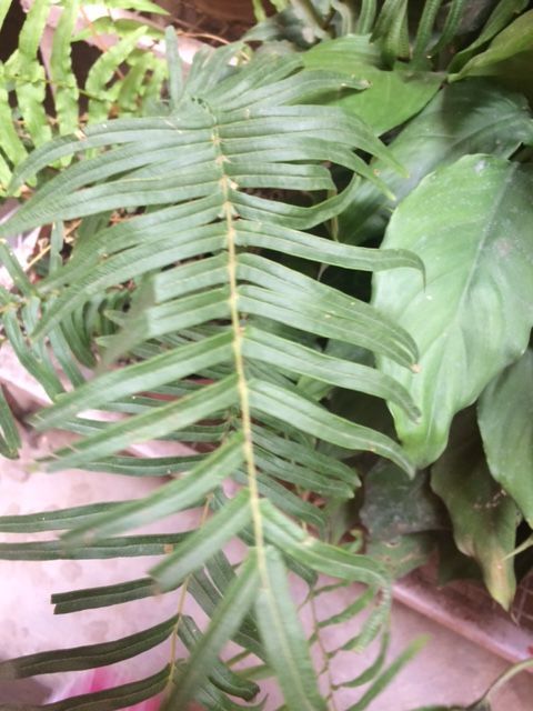
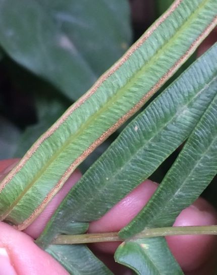
Our first practical material to begin was the fern Pteris vittata , growing in the college Botanical garden. The fertile leaves were bearing sporangia along the ventral margins. We took out some sporangia and prepared a temporary wet mount. After fitting the slide in the foldscope, the first observations were clicked using a simple phone.
It was a great honor to be selected by Foldscope team for use of Foldscopes as a teaching and educational tool. The orientation workshop at ICGEB, New Delhi on 16 th April, 2018 gave us an opportunity to interact with our N-E twining parteners. Learning the assembly of Foldscopes from Prof. Jim and Dr. Shailja’s team at ICGEB marked the beginning of the project.
My 1 st hand experience with the foldscope
I am excited to share the results of our 1 st attempt of using Foldscope along with my Co-PI Dr. Jasveen Dua
After the orientation at ICGEB, New Delhi on assembly and usage of Foldscope, the few initial days at Chandigarh passed completing the documentation and other formalities to start working with the Foldscopes.
Our first practical material to begin was the fern Pteris vittata , growing in the college Botanical garden. The fertile leaves were bearing sporangia along the ventral margins. We took out some sporangia and prepared a temporary wet mount. After fitting the slide in the foldscope, the first observations were clicked using a simple phone.
View in Media Gallery
Fern Pteris vittata
View in Media Gallery
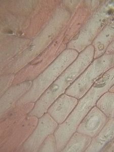
Pteris Leaf
View in Media Gallery
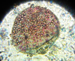
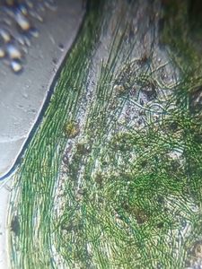
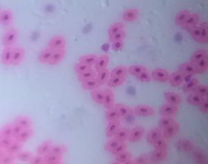
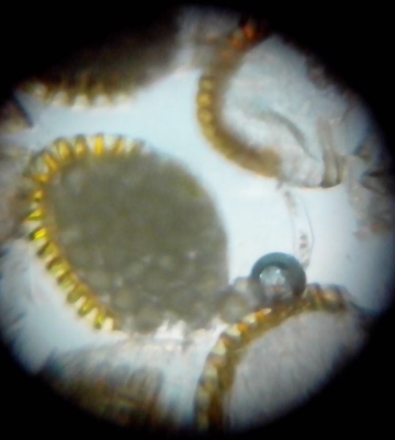
A group of sporangia filled with spores
View in Media Gallery
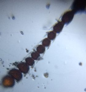
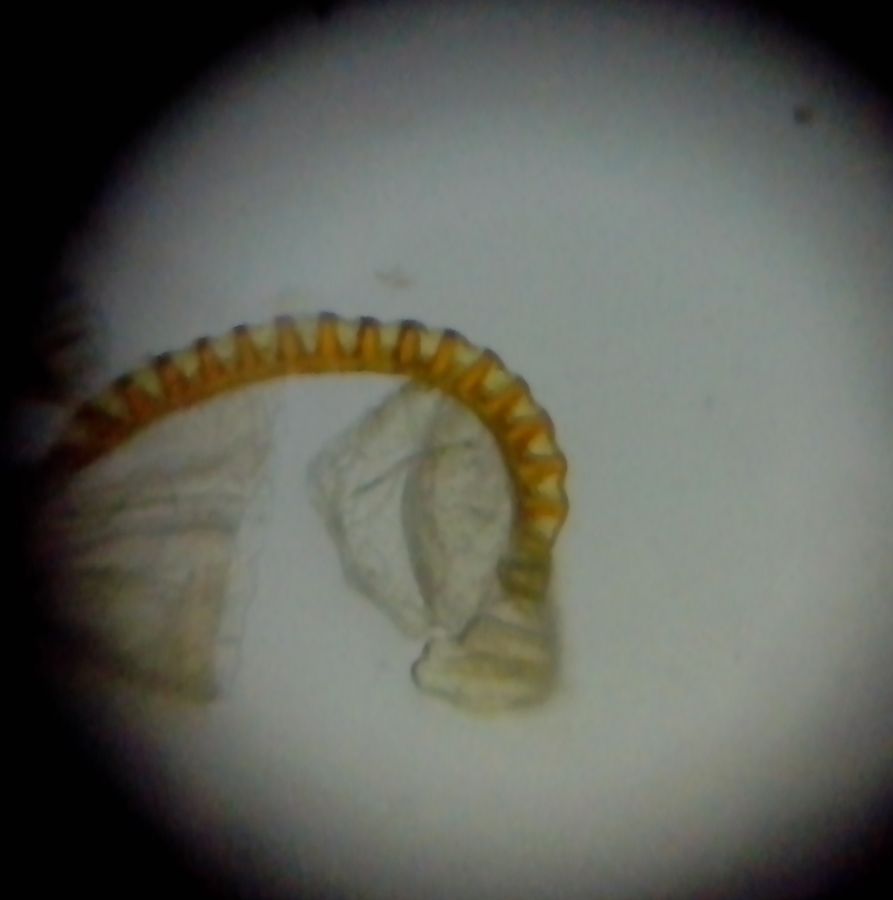
Each sporangium had a long stalk and an outer boundary of cells with thickened radial walls. Some triangular shaped spores with thick outer covering
View in Media Gallery
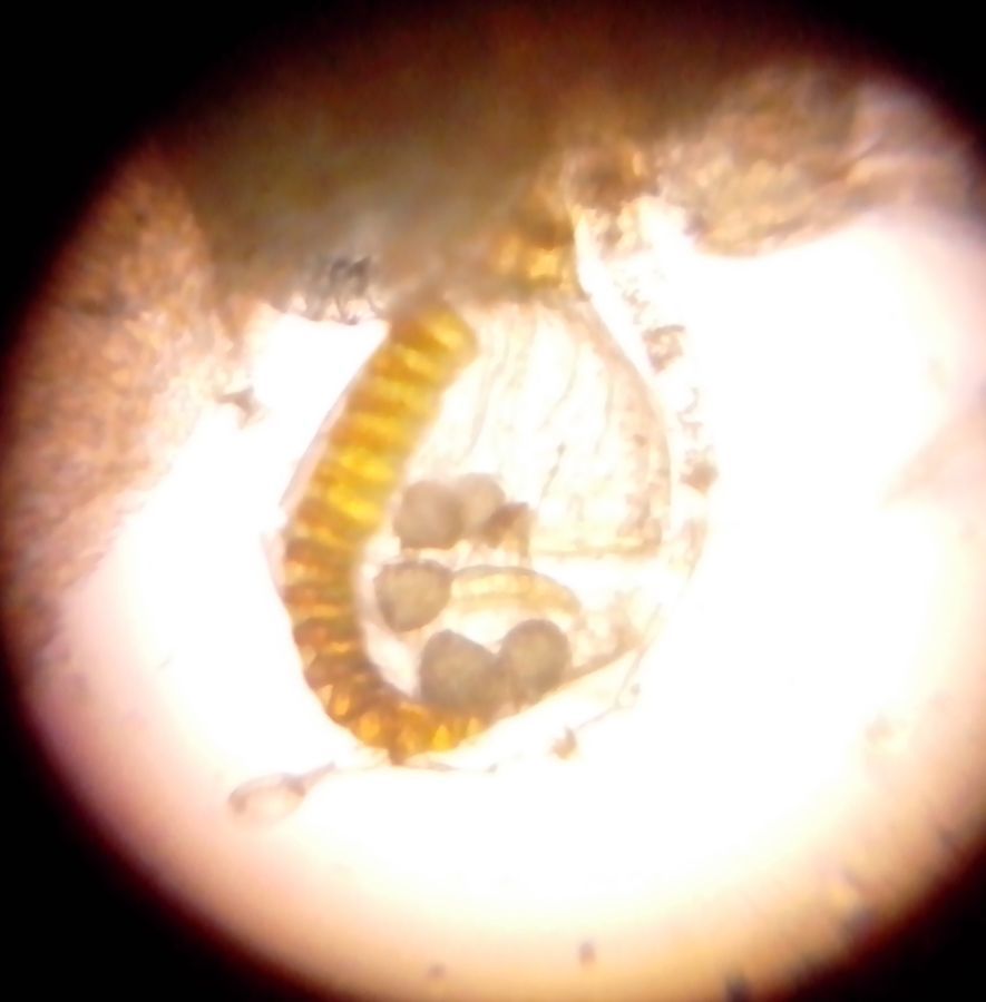
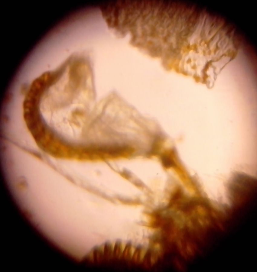
Sporangium opening with released spores
View in Media Gallery
A few thin-walled cells from where spores were released marked the point of opening of sporangium
Sign in to commentNobody has commented yet... Share your thoughts with the author and start the discussion!

 0 Applause
 0 Comments