Imaging of Onion Peel Cells Through Foldscope
Jul 18, 2018 • 6:15 AM UTC
Unknown Location
140x Magnification
Microorganisms
Jitendra Satija
Learn about the author...
8
posts
6
comments
0
locations
0 Applause
0 Comments
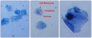
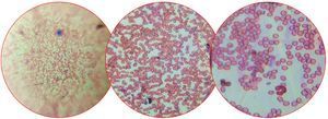
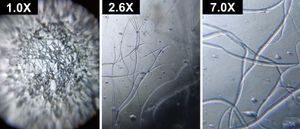
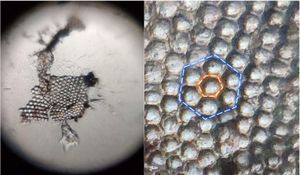
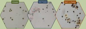
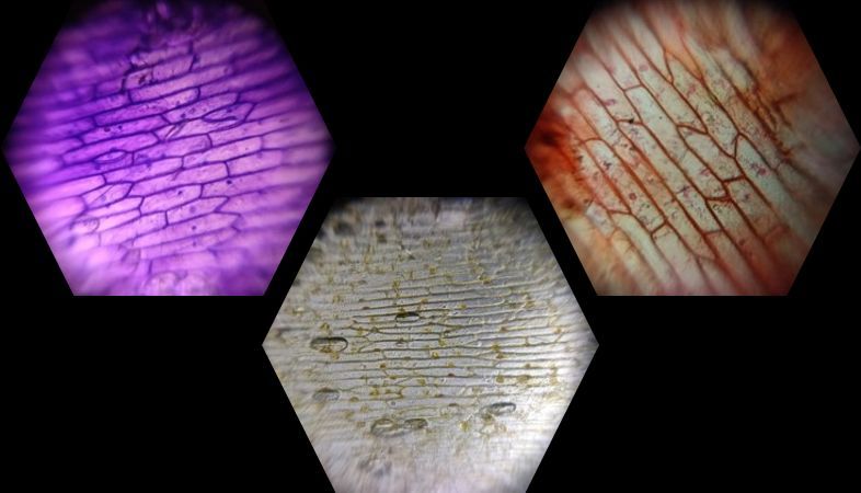
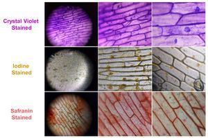
Onion peel cells were stained using crystal violet, iodine, and safranin dye separately and imaged under foldscope.
Sign in to comment
Nobody has commented yet... Share your thoughts with the author and start the discussion!
More Posts from Jitendra Satija
Foldscope: Hands On Workshop cum Image Acquisition Contest
0 Applause
0 Comments
6y
Observation of Methylene Blue-stained Cheek Cells using Foldscope
0 Applause
0 Comments
6y
Observation of Stained Blood Smear Using Foldscope
0 Applause
0 Comments
6y
Foldscope application in quality control of electrospun fibers by observing beads formation
0 Applause
0 Comments
6y
Hexagonal Array Packing of Ommatidia in Mosquito Eye through Foldscope
0 Applause
0 Comments
6y
Rotifers: Beating of their Ciliary Crown, Feeding and Locomotory Behaviour through Foldscope
0 Applause
0 Comments
6y
Microscopic Characterization of Salt-Stress Induced Cell Death in the Unicellular Green Algae Chlorella vulgaris
0 Applause
0 Comments
6y