HeLa Cell staining by plant dye and observation using Foldscope
 Nov 01, 2018 • 9:49 AM UTC
 Unknown Location
 140x Magnification
 Microorganisms
ANUTOSH PATRA
Learn about the author...
6posts
0comments
1locations

View in Media Gallery
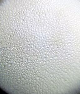
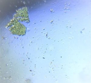
HeLa cells are cultured on the glass slide coated with 2% gelatin Washed cells with 1X PBS, stained with plant dye Observed under Foldscope and conventional microscope for comparison A- Unstained cells. Under conventional microscope with 200 X zoom B-Stained cells, under the conventional microscope with 200 X zoom
C- Unstained cells. Under Foldscope, photography captured by 12 MP smartphone camera with 2X digital zoom
D- Stained cells, Under Foldscope, photography captured by 12 MP smartphone camera with 2X digital zoom
Observed under Foldscope and conventional microscope.
C- Unstained cells. Under Foldscope, photography captured by 12 MP smartphone camera with 2X digital zoom
D- Stained cells, Under Foldscope, photography captured by 12 MP smartphone camera with 2X digital zoom
Observed under Foldscope and conventional microscope.
View in Media Gallery
Sign in to commentNobody has commented yet... Share your thoughts with the author and start the discussion!

 0 Applause
 0 Comments