Poison oak wounds on my hands
 Sep 10, 2015 • 6:55 PM UTC
 Unknown Location
 140x Magnification
 Microorganisms
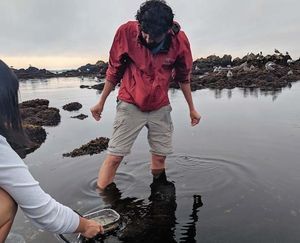
Manu Prakash
I am a faculty at Stanford and run the Prakash Lab at Department of Bioengineering at Stanford University. Foldscope community is at the heart of our Frugal Science movement - and I can not tell you how proud I am of this community and grassroots movement. Find our work here: http://prakashlab.stanford.edu
266posts
1198comments
42locations

View in Media Gallery
This is probably the most painful story I will share (I hope I don’t have to go through it again). But for the sake of science, we sometimes do put ourselves in awkward and uncomfortable situations. So here I go..
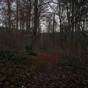
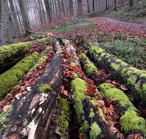
Recently, during a late night field work at a new site we had never worked before; I ended up guiding my whole research group through a minefield of “poison oak”. See, I grew up in India, and had only heard about poison oak from friends (and I did not believe them). The field work was quiet successful actually and we continued working at this site for the next three days.
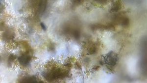
But after two weeks, almost all of us started getting blisters and the whole deal. I don’t have to describe to you what happens in poison oak reaction; but let me jus quickly share data from the wound sites itself.
Tutorial:
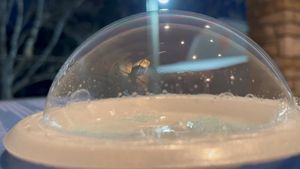
Since I have not described how to use Foldscope to image skin surface before, here is a quick hint. You need to remove the light module completely. Unfold the portion which holds the light module as well; this exposing a major portion of the lens. Now bring it close to skin, and turn on a table lamp pointing to your arm close by. You will see the light leaking from the side, bouncing from surface and getting to your camera. That’s it.
Here is a video , where I image my skin surface using a reflected light microscopy trick.
Recently, during a late night field work at a new site we had never worked before; I ended up guiding my whole research group through a minefield of “poison oak”. See, I grew up in India, and had only heard about poison oak from friends (and I did not believe them). The field work was quiet successful actually and we continued working at this site for the next three days.
But after two weeks, almost all of us started getting blisters and the whole deal. I don’t have to describe to you what happens in poison oak reaction; but let me jus quickly share data from the wound sites itself.
Tutorial:
Since I have not described how to use Foldscope to image skin surface before, here is a quick hint. You need to remove the light module completely. Unfold the portion which holds the light module as well; this exposing a major portion of the lens. Now bring it close to skin, and turn on a table lamp pointing to your arm close by. You will see the light leaking from the side, bouncing from surface and getting to your camera. That’s it.
Here is a video , where I image my skin surface using a reflected light microscopy trick.
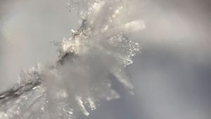
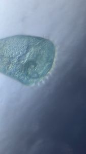
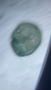
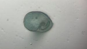
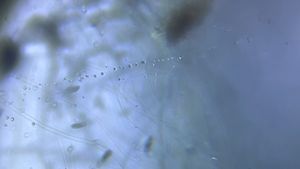
The blisters and wounds look like volcano. You want to watch the whole video to find the sections with healing blisters.
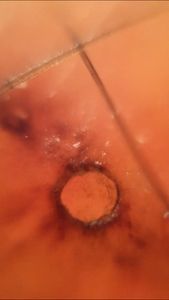
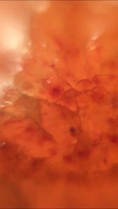
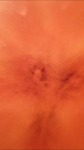
It felt very strange watching this – since this was my own skin. Also, watching the healing and small pockets of activity (red areas) made me realize that a wound site is almost like a war zone. Extreme amount of activity that happens. Next time, I am going to do a time lapse of an injury site.
Here is an experiment I don’t want to repeat.
Cheers
Manu
Here is an experiment I don’t want to repeat.
Cheers
Manu
Sign in to commentNobody has commented yet... Share your thoughts with the author and start the discussion!
 0 Applause
 0 Comments