LGP26 B-2 Day 3 Rahul
 May 28, 2026 • 1:21 PM UTC
 India
India 340x Magnification
 Plants
Rahul
Learn about the author...
6posts
0comments
0locations
View in Media Gallery
Hi everyone,
Today was my third day using the Foldscope. During my time, I observed multiple specimens including Hibiscus pollen, Sunflower stem and Sunflower pollen. Below are my results and observations.
Today was my third day using the Foldscope. During my time, I observed multiple specimens including Hibiscus pollen, Sunflower stem and Sunflower pollen. Below are my results and observations.

View in Media Gallery
This is the Hibiscus flower which I have examined. From the outside, it is pink in color and has yellow pollen. The petals are very soft. Now let's move inside the pollen.

View in Media Gallery
This is the 50x magnification of the pollen. The Hibiscus flower was take outside the residence hall on the campus. It was found within a garden. The ovaries of the flower (this point is for all flowers) is the pollen or fruit. Thus, we have been observing the ovaries of the flower. In the image, I can see yellow cells with slightly black spots. However, the cells are too small to view the cell effectively.

View in Media Gallery
This is the 140x magnification of the sample. The camera was slightly blurry, however the slide was good enough to make accurate observations. As you can see, the pollen doesn't have to be circular. There are some irregular pollen (oval shaped). Also, the spikes on the membrane of the pollen is visible. Still, not many internal parts are viewable.

View in Media Gallery
This is the final magnification of the Hibiscus flower at 340x. The image is still blurry due to the camera. However, the spikes have more clarity as they can be viewed with the blur. The yellow color remains throughout each magnification. We still cannot see inside the pollen, showing how small the pollen particles really are.

View in Media Gallery
This is the sunflower pollen which is observed at 50x. The particles are very minute and difficult to view, especially at 50x. However, it can be seen that the cells are yellow, but no internal details are visible.
The 140x was very blurry, thus I was unable to post it. The only difference was that the cells were larger. There was no other change.
The 140x was very blurry, thus I was unable to post it. The only difference was that the cells were larger. There was no other change.

View in Media Gallery
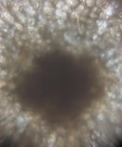
This is diagram representing the sunflower pollen at 340x. There is much clarity. This is because as you can see, the spikes are very visible. However, the color changed slightly becoming darker.

View in Media Gallery
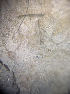
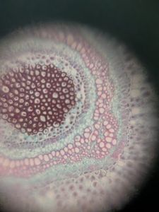
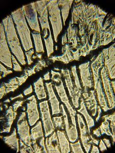
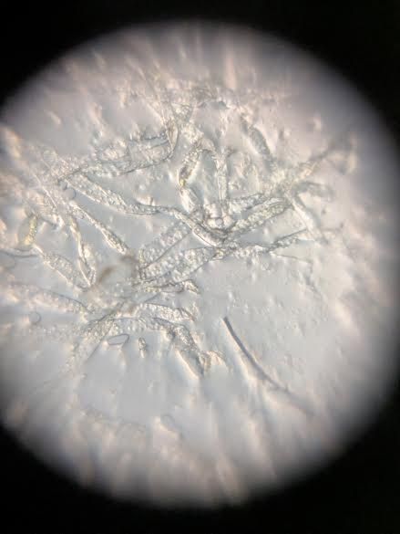
This is the sunflower stem or fiber. This was very difficult to observe. It has a neutral color and is very elongated. This was only done at 140x due to time restrictions.
-Rahul
-Rahul
Sign in to commentNobody has commented yet... Share your thoughts with the author and start the discussion!
 0 Applause
 0 Comments