LGP 26 B-2 Day 4 Rahul
 May 28, 2026 • 2:17 PM UTC
 India
India 340x Magnification
 Plants
Rahul
Learn about the author...
6posts
0comments
0locations
View in Media Gallery
Hi everyone,
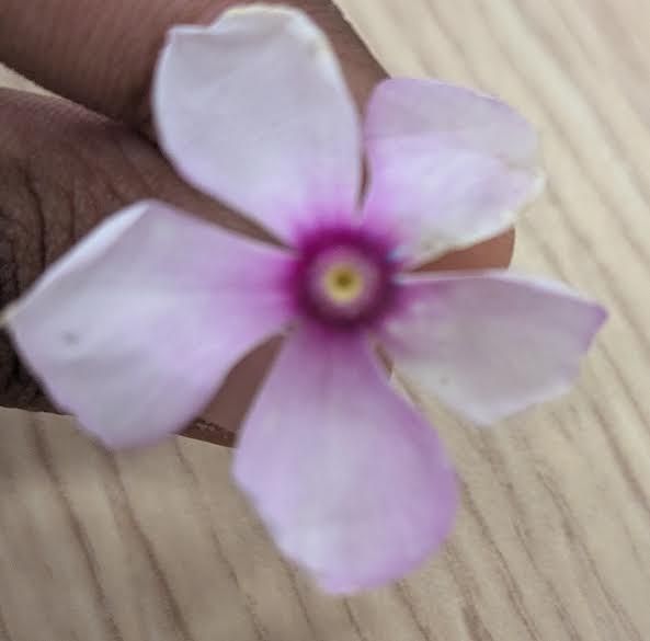
Today was Day 4 of Exploring The Microcosm in Lodha Genius Program at Ashoka University. It was an amazing time where I observed Periwinkle, Pumpkin Pollen, and Leaf Fiber and Stomata.
Today was Day 4 of Exploring The Microcosm in Lodha Genius Program at Ashoka University. It was an amazing time where I observed Periwinkle, Pumpkin Pollen, and Leaf Fiber and Stomata.

View in Media Gallery
This is a sample of periwinkle at 50x magnification. As you can see, it is small black bean-like structures. It is very difficult to observe as it is so small. The image is blurry even on the slide, indicating that we should increase the magnification.

View in Media Gallery
This is the same sample at 140x magnification. The structure is being more visible. I can notice the large cluster of periwinkle pollen. Also, at this point, you can begin to notice the slight neutral color inside the pollen.

View in Media Gallery
This is the 340x magnification of the periwinkle sample. As you can see, an increase the magnification improved the clarity of the image/slide. Additionally, it is much easier to see the neutral color within the cell. There is a black outline on the cell. In the picture, the cells are very crowded (maybe I took too many samples).

View in Media Gallery
This is the 50x magnification of the Pumpkin Pollen. In the picture, I can see 2 yellow pollens which are surrounded by air molecules. The pollen is bigger than other pollen that were previously viewed, but it is too small to see internal parts. Additionally, as shown, the color of the pollen is distinctly yellow.

View in Media Gallery
This is the 140x magnification of the Pumpkin Pollen. With the increase in magnification, there is an increase in visibility of the image, and thus a image with more image is generated. Moreover, there are some black spots indicating pollen parts visible in this image.

View in Media Gallery
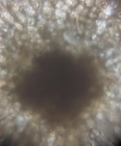
This is the 340x magnification of the Pumpkin Pollen. While the quality of the image was terrible, the slide viewing was very well. The clarity allowed us to see so much detail such as the hair (not really spikes) that were on the pollen. However, again, I think I could way too much sample.

View in Media Gallery
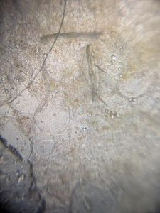
This is the 50x magnification of the Leaf Stem/Fiber. The image is blurry, thus we need to increase the magnification. However, we can notice that it is black cells, however I believe their color will change when we zoom in.

View in Media Gallery
This is the 140x magnification of the Leaf Stem/Fiber. At this magnification, the image becomes clearer, but I think we need to increase the magnification further to receive an accurate photo. The neutral color of the pollen is starting to become visible.

View in Media Gallery
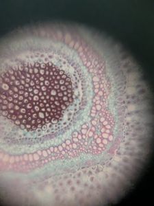
This is the 340x magnification of the Leaf Stem/Fiber. Finally, the clarity of the image is perfect. This shows that sometimes your image is blurry due to low magnification (this occurs in the stomata as well). The fiber has a black outline with a neutral inside and the structure is very elongated.

View in Media Gallery
This is the 50x magnification of the Leaf Stomata. As we can see, the same clarity problem is occurring, thus we will increase magnification. However, you can notice a little bit of green.

View in Media Gallery
This is the 140x magnification of the Leaf Stomata. The clarity problem continues. Now, the green is more visible which is caused by the chloroplasts in the non-stomata cells.

View in Media Gallery
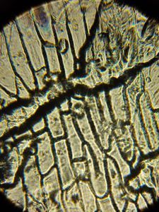
This is the 340x magnification of the Leaf Stomata. Again, at the final magnification, the image becomes clear. The stomata can be seen opening and closing to transpire. Thus, the white lines are the openings and the stomata are the boundary cells of a plant.
-Rahul
-Rahul
Sign in to commentNobody has commented yet... Share your thoughts with the author and start the discussion!
 0 Applause
 0 Comments