Parasitic infections in Cattle (a. Cattle ticks)
 Sep 30, 2018 • 10:53 PM UTC
 Unknown Location
 140x Magnification
 Unknown
Meignanalakshmi Sundaram
I am Dr.S.Meignanalakshmi, working as Professor, at the Directorate of Centre for Animal Health Studies, TANUVAS, Chennai-51. Working on Foldscope project on "Foldscope for diagnosis of Rumen Acidosis and parasitic infections in cattle" sanctioned by DBT
66posts
8comments
1locations
View in Media Gallery
Endoparasites live inside the host. The may either be microparasites such as: blood protozoans (Theileria, Babesia, Trypanosomes) or macroparasites like: Helminths (Trematodes, Cestodes, Nematodes)
Ectoparasites include ticks, mites, leeches, flies etc . They live on the outer surface of the host. The most common ectoparasites are ticks which causes cattle tick fever, Theileriosis ,Babesiosis etc.
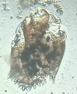
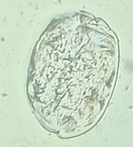
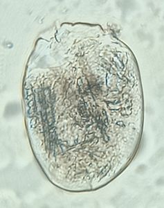
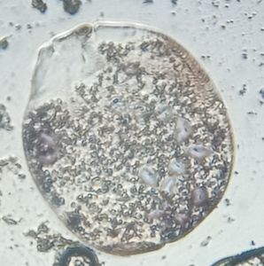
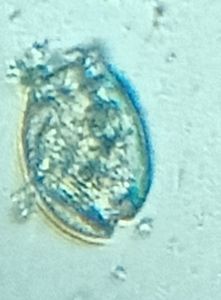
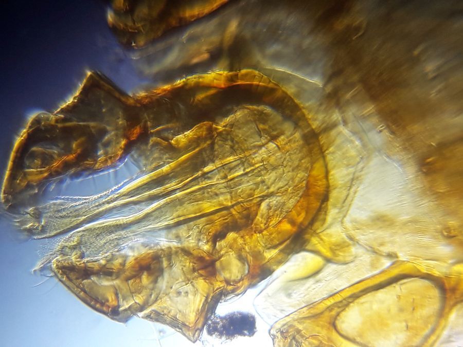
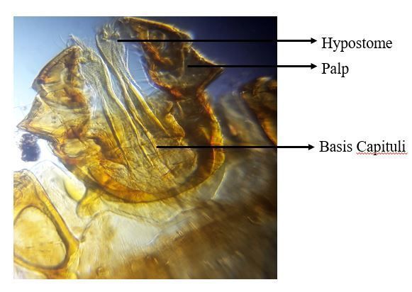
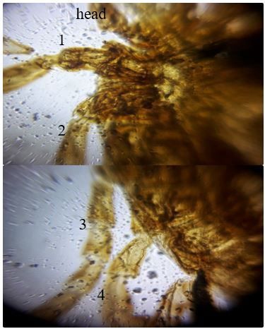
Ticks were collected from a cow suspected of Theileriosis showing the following symptoms: fever, tick infestation, rough coat, restlessness, decreased milk production etc. and stored in 10% Formalin and basic fixation techniques were carried out. Based on the Anatomy, the tick was identified as Boophilus microplus in the Nymph stage (8 legs). Various parts were identified under a foldscope.
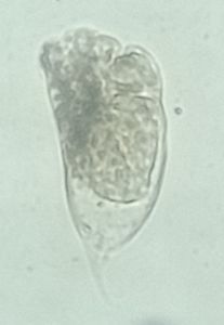
Head
Ectoparasites include ticks, mites, leeches, flies etc . They live on the outer surface of the host. The most common ectoparasites are ticks which causes cattle tick fever, Theileriosis ,Babesiosis etc.
Ticks were collected from a cow suspected of Theileriosis showing the following symptoms: fever, tick infestation, rough coat, restlessness, decreased milk production etc. and stored in 10% Formalin and basic fixation techniques were carried out. Based on the Anatomy, the tick was identified as Boophilus microplus in the Nymph stage (8 legs). Various parts were identified under a foldscope.
Head
View in Media Gallery
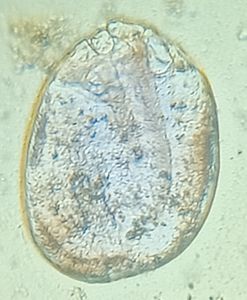
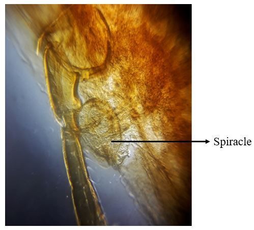
2. Spiracle
View in Media Gallery
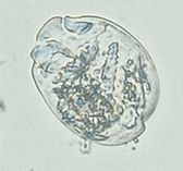
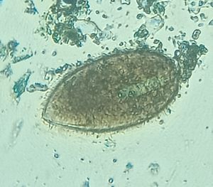
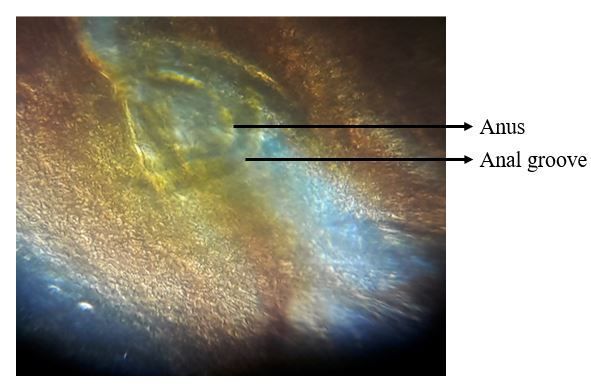
3. Anus and anal groove.
View in Media Gallery
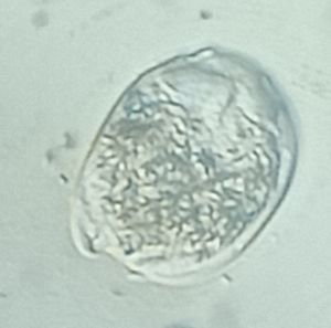
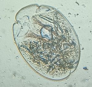
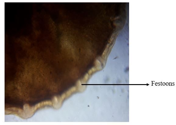
5. Festoons
They are small areas separated by short grooves on the back margin of the tick. This helps to distinguish all other ticks from Ixodes ticks, which lack festoons.
They are small areas separated by short grooves on the back margin of the tick. This helps to distinguish all other ticks from Ixodes ticks, which lack festoons.
View in Media Gallery
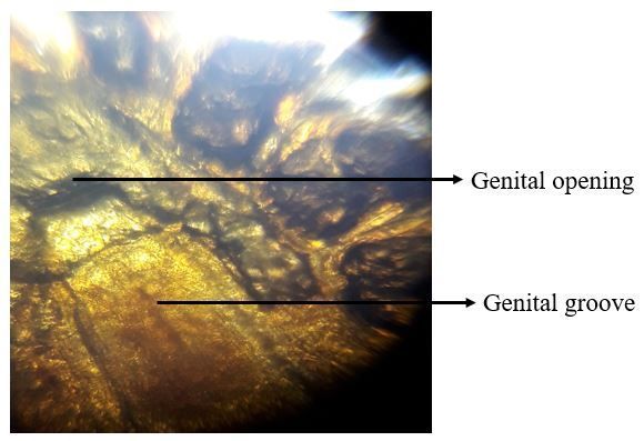
6. Genital opening and groove
View in Media Gallery
7. 4 legs indicating that larvae is in nymph stage
View in Media Gallery
Sign in to commentNobody has commented yet... Share your thoughts with the author and start the discussion!

 0 Applause
 0 Comments