Exploring the Microcosm Day 1 Part 2 #LGP26-B3
 Jun 02, 2026 • 7:07 PM UTC
 India
India 140x Magnification
 Plants
Moucham
Learn about the author...
6posts
0comments
0locations
View in Media Gallery
On the first day of our hands on week long course “Exploring the Microcosm”, we observed Fern Rhizomes (The blog post for the same can be found in my profile) and then onion peels. We prepared a glass slide to observe the peel of onion. We observed the cell under magnifications of 50x, 140x and 340x.

View in Media Gallery
The above is at 50x magnification and gives us a wider spectrum of the onion cells and is arranged in an organized brick-like pattern. They are closely packed in parallel rows with very less intercellular space between them. Here, we can infer that individual cells appear elongated and rectangular. We can clearly observe distinct vertical and horizontal lines separating each cell. These horizontal and vertical separations are nothing else but the protective cell walls made of cellulose, which gives the respective cells their fixed shape. At this magnification, the cell organelles aren’t visible.

View in Media Gallery
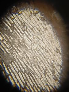
The above is at 140x and thus gives a magnified image of the onion cell and now as the field of view is much narrower, instead of seeing a massive grid of cells you get a few individual cells to focus upon. The outlines represent a closer look at the cell walls, showing a distinct demarcation between 2 adjacent cells. The central cell in focus shows the conventional rectangle, tube like shape structure and are characteristics of onion epidermal tissue.

View in Media Gallery
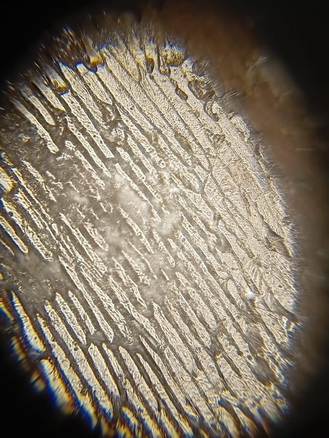
The above is at 340x magnification of the onion cell. This magnification is large enough such that you aren’t looking at bundle of cells but few individual cells. The long tracks of lines going across the field of view show a “double line” structure. This is a highly magnified view of cell walls, separating adjacent cells from each other.
With these observations in mind, we can conclude day one of the course “Exploring the Microcosm”.
With these observations in mind, we can conclude day one of the course “Exploring the Microcosm”.
Sign in to commentNobody has commented yet... Share your thoughts with the author and start the discussion!

 0 Applause
 0 Comments