Exploring the Microcosm #LGP_26-B3
 Jun 03, 2026 • 1:04 PM UTC
 India
India 140x Magnification
 Plants
Moucham
Learn about the author...
6posts
0comments
0locations
View in Media Gallery
After having analyzed Onion cells and Fern Rhizomes cells on day 1 of the course “Exploring the Microcosm”, we moved into the second day of the course with the same excitement and rather more curiosity than the previous day. On the 2nd day of the abovementioned course at the Lodha Genius-Ashoka University Programme, we viewed potato peel and potato flesh samples under the foldscope. We viewed these samples on the foldscope after preparing the sample on the glass slide.

View in Media Gallery
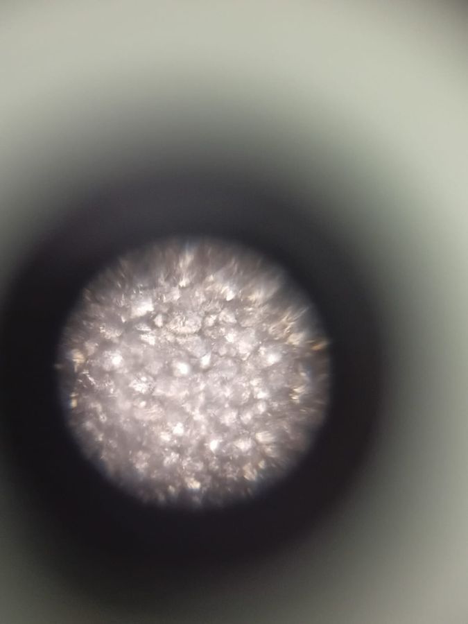
The above is the image of a potato flesh cell viewed under the foldscope at a 50x magnification, and some critical observations can be inferred from the same. If we look closely at the faint, we can find overlapping boundaries within the bright circular image of the cell. The most visible component of the image above is the dense and translucent crystalline structures which are possibly the leucoplast, which are non-pigmented and are specialized cells. They are responsible for store starch. The reason the centre is highly laminating is that the light source was directly behind the sample.

View in Media Gallery
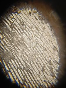
The above image is the same sample of potato flesh but under a 140x lens, and this image gives us few more insights upon the nature and composition of the cell. At 50x magnification, the centre looked like a singular mass, but at 140x the image breakdowns into distinct and individual components, we can view individual starch grains clustered inside the cells with very dense composition. At the upper region of the image, you can make a trivial observation that the grains aren’t perfectly round, which I believe is because they are compressed tightly against each other thus giving an irregular or faceted shape to them. As previously observed, there are molecules which are tightly packed into these membrane bound organelles. This image shows how the starch is stored with almost zero space between them.

View in Media Gallery
The above image we are viewing is the potato flesh cells at 340x. At 50x and 140x we were looking at cell clusters but at 340x the field of view has narrowed deeper into the tissues to an extend that those boundaries which looked like cobblestones can be viewed as massive, individual starch granules or just few tight clusters of granules.
In conclusion, we could make various interpretations upon our images we received from the foldscope.
In conclusion, we could make various interpretations upon our images we received from the foldscope.
Sign in to commentNobody has commented yet... Share your thoughts with the author and start the discussion!

 0 Applause
 0 Comments