Day 1 with the foldscope
 Jun 02, 2026 • 10:29 AM UTC
 India
India 140x Magnification
 Plants
Moucham
Learn about the author...
6posts
0comments
0locations

View in Media Gallery
Exploring the Microcosm #LGP26-B3 Day 1 At the first seminar lecture of the course “Exploring the Microcosm”, we observed Fern Rhizomes under the foldscope, having made various observations under different lenses which are 50x, 140x and 340x. While initially it turned out difficult to get clear pictures of the images, it turned out to be extremely fun moving forward. Using the foldscope for the first time was indeed enriching.
We started off observing Fern Rhizomes. We observed it using three distinct lenses, 50x, 140x and 340x and different images/observations could be made.
We started off observing Fern Rhizomes. We observed it using three distinct lenses, 50x, 140x and 340x and different images/observations could be made.

View in Media Gallery
The above is Fern Rhizome at 340x magnification, and some trivial and clear observations can be made. We can observe the cell walls and also the nucleus of the cell. The cells are in a hexagonal shape.

View in Media Gallery
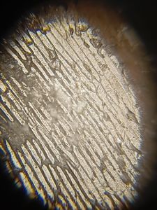
The above is Fern Rhizome at 140x magnification, and again we can make some distinct observations and at the same time make distinctions between the 340x magnification image. We can observe the pink-ish hexagonal structures of the cell image we observed in the 340x magnification, with other distinct components of the cells which were previously not visible.

View in Media Gallery
The above is Fern Rhizome at 50x magnification, and again we can see visible differences between this picture and previous ones under lenses of different magnifications. Although some parts of this turned out to be blurry, we can still make our interpretations.
All of these observations were made under a pre-stained glass slide which was stained by Fern Rhizome.
All of these observations were made under a pre-stained glass slide which was stained by Fern Rhizome.
Sign in to commentNobody has commented yet... Share your thoughts with the author and start the discussion!

 0 Applause
 0 Comments