Microcosm Day 3 #LGP26_B3
 Jun 04, 2026 • 5:28 PM UTC
 India
India 140x Magnification
 Plants
Moucham
Learn about the author...
6posts
0comments
0locations
View in Media Gallery
The 3rd day of our workshop “Exploring the Microcosm” started off in high spirits as it did on the first two days. After having observed Onion Cells and Potato Cells on the very day, we observed tomato cells and pollen on the 3rd day of the workshop. We first started off with observing tomato peels and then observed the flesh of the tomato, both of which gave me results which I reflected upon.
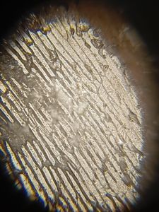
Let’s start off noting our observations for tomato peel at various magnifications. At 50x magnification, the foldscope provides a wide and comprehensive view of the epidermal tissue layer. At a glance, we can intuitively infer that the cells are continuous and are tightly packed with very little to no intercellular space between them. We can observe an uneven yet prominent and vibrant colour distribution of orange, yellow and some reddish shades, visible across the layer. We can also look at the visible discontinuation between the red and white layer, where the white layer basically is the mounting medium on the glass slide. At 140x magnification, the field of view narrows significantly further. We can view contours of individual cells in focus. The shapes exhibit irregular or polygonal shape. The cells also give out a granular appearance. This is probably caused due to tightly packed components. The variations in the colour of cells become clearer and distinct, showing localized concentrations of yellow and deep orange pigments within cell boundaries of individual cells.
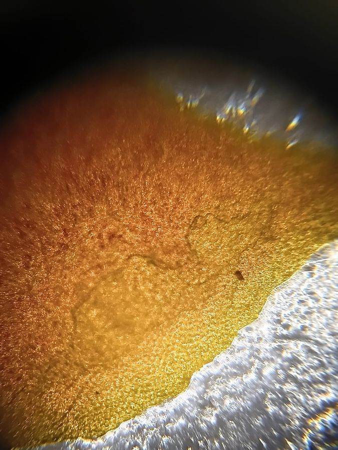
Going forward we also analyzed Tomato flesh cells under various magnifications. At 50x magnification, we get a broad layout overview of the tissue. The cells appear as a dense, continuous mass of large, loosely packed structures. The boundaries between distinct cells are visible but not detailed. The cells we observe are mostly spherical. A faint, uniform reddish-orange or yellowish tint is visible across the field of view. Next we viewed them under 140x magnification. This magnification bridges the gap, allowing you to focus on the structure of individual cells. At this magnification, the thin primary cell walls of the parenchyma cells become distinctly visible. Small, granular dots or needle-like structures with a distinct red/orange color become visible within the cytoplasm. These are chromoplasts. We can view small intercellular spaces in the corners where several spherical cells meet. We further moved into 340x magnification. The reddish orange chromoplasts can be viewed under a higher definition. With careful adjustment of the fine focus, a faint, grainy, spherical nucleus may be visible, typically pushed against the cell wall due to the pressure of the large vacuole.
Let’s start off noting our observations for tomato peel at various magnifications. At 50x magnification, the foldscope provides a wide and comprehensive view of the epidermal tissue layer. At a glance, we can intuitively infer that the cells are continuous and are tightly packed with very little to no intercellular space between them. We can observe an uneven yet prominent and vibrant colour distribution of orange, yellow and some reddish shades, visible across the layer. We can also look at the visible discontinuation between the red and white layer, where the white layer basically is the mounting medium on the glass slide. At 140x magnification, the field of view narrows significantly further. We can view contours of individual cells in focus. The shapes exhibit irregular or polygonal shape. The cells also give out a granular appearance. This is probably caused due to tightly packed components. The variations in the colour of cells become clearer and distinct, showing localized concentrations of yellow and deep orange pigments within cell boundaries of individual cells.
Going forward we also analyzed Tomato flesh cells under various magnifications. At 50x magnification, we get a broad layout overview of the tissue. The cells appear as a dense, continuous mass of large, loosely packed structures. The boundaries between distinct cells are visible but not detailed. The cells we observe are mostly spherical. A faint, uniform reddish-orange or yellowish tint is visible across the field of view. Next we viewed them under 140x magnification. This magnification bridges the gap, allowing you to focus on the structure of individual cells. At this magnification, the thin primary cell walls of the parenchyma cells become distinctly visible. Small, granular dots or needle-like structures with a distinct red/orange color become visible within the cytoplasm. These are chromoplasts. We can view small intercellular spaces in the corners where several spherical cells meet. We further moved into 340x magnification. The reddish orange chromoplasts can be viewed under a higher definition. With careful adjustment of the fine focus, a faint, grainy, spherical nucleus may be visible, typically pushed against the cell wall due to the pressure of the large vacuole.


Sign in to commentNobody has commented yet... Share your thoughts with the author and start the discussion!

 0 Applause
 0 Comments