Exploring the Microcosm Day 2 Part 2 #LGP26-B3
 Jun 04, 2026 • 7:16 AM UTC
 India
India 140x Magnification
 Plants
Moucham
Learn about the author...
6posts
0comments
0locations
View in Media Gallery
With great excitement, we started off the second day of our week long workshop titled “Exploring the Microcosm” at the Lodha Genius- Ashoka University Programme. On the second day, we viewed onion peel cells and onion flesh cells on the foldscope, with the observations on the potato flesh cells detailed in my previous blog.

View in Media Gallery
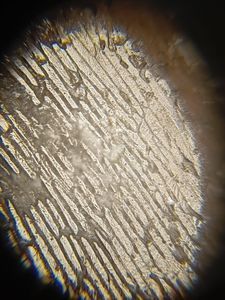
The above image is the peel of the potato at 50x magnification. The tissue layout here is completely different from the tissue layout of the tissues in the potato peel. If you look at the upper and outer edges of the illuminated circle, the texture of the cells change from large, loose shapes we saw in the potato flesh to denser and very tightly packed. Unlike the interior potato flesh cells which are designed to expand and store starch, these outer cells are dead maturity, highly compacted and arranged in orderly, overlapping rows. The job of these peel cells is to act as tough structural shields for the other components and thus are dead to reduce energy consumption. The individual glister of cells which we saw in the flesh cells. The centre is more diffused and uniform.

View in Media Gallery
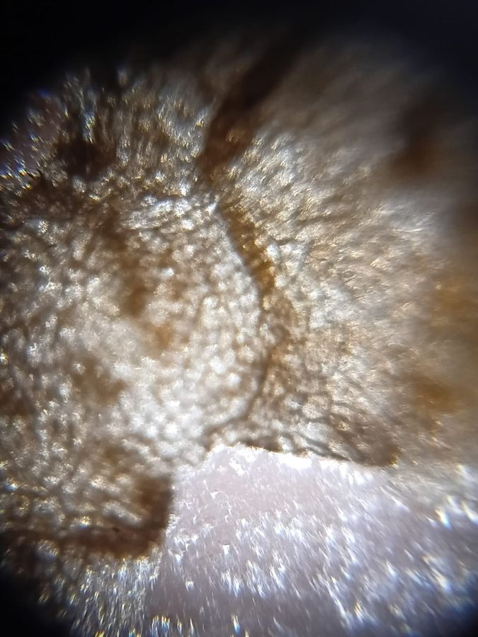
The above image is the peel of the potato at 140x magnification. This gives a little more rigidness and layout compared to the cell we viewed at 50x magnification. The texture has expanded into massive network of tightly packed cells. You can see distinct borders tracing around numerous individual cellular units. There are also deep, brown wavy bands and patches cutting through the field of view. These probably represent dense concentrations of suberin and tannins embedded in the cell layers.

View in Media Gallery
The above is the image of the peel of the potato at 340x magnification. At such a magnification ie 340x you can view distinct geometric boundaries where the individual granules meet. The left and center-left areas display a highly organized, geometric grid of rectangular, boxy cells. This is the view of the cork layer. These cells are dead at maturity, fully devoid of cytoplasm and act strictly as a protective layer to protect the cells soil pathogens, insects and physical damage. The cells in the exact centre can be viewed distinctly and are incredibly sharp, while the cellular grid begins to compress into blurred images at the bottom edge of the frame.
Sign in to commentNobody has commented yet... Share your thoughts with the author and start the discussion!

 0 Applause
 0 Comments