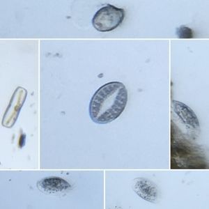
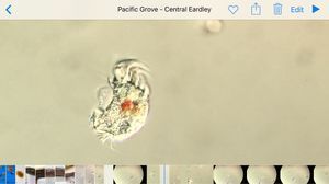
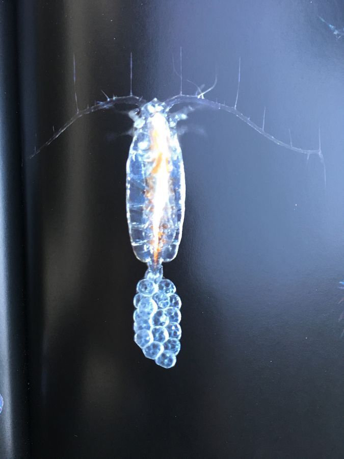
Copepod egg sac
 Nov 12, 2015 • 12:39 AM UTC
 Unknown Location
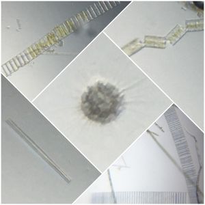
 140x Magnification
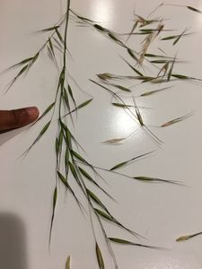
 Plants
Saad Bhamla
Learn about the author...
32posts
11comments
2locations

View in Media Gallery
This is a meta learning post of how the foldscope contributes to my learning and growth, which I felt was worth sharing.
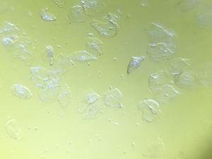
From the same Hopkins trip where I found the nudibranch eggs, I also was able to find Copepods swimming in the ocean water sample.
I took two videos exactly and couldn’t make head or tail (literally) of the Copepods. I left the videos on my phone and forgot about them, until right now. I’m sure you have a similar experience where you stick something under the foldscope but don’t ‘learn’ anything per se from the experiment.
Today, I came across this book from the stanford library and I was leafing through it and saw the following images.

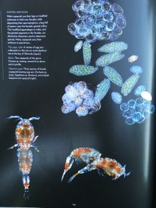
Page 148 describes Copepods and the caption says that female Copepods can be distinguished by the presence of egg sacs near the tail. And there is the following image on page 149.
View in Media Gallery
As soon as I saw these images, a bulb went off in my brain. I recalled seeing something similar. With my own eyes !! So I pulled out my phone and was able to find the following two videos which in my mind I has written off as ‘ meh, not interesting ‘, perhaps because I was just exhausted that day after hours of foldscoping 🙂


So, clearly what I was observing were the beautiful egg sacs on a female copepod.
The lesson here, at least for me, is that sometimes it’s easy to think that just because you see through a foldscope, you’ll learn instantly or not learn something. Sometimes it takes time to make the connections.
So, be patient, and keep exploring.
-Saad
The lesson here, at least for me, is that sometimes it’s easy to think that just because you see through a foldscope, you’ll learn instantly or not learn something. Sometimes it takes time to make the connections.
So, be patient, and keep exploring.
-Saad
Sign in to commentNobody has commented yet... Share your thoughts with the author and start the discussion!

 0 Applause
 0 Comments