Detecting Hepatitis B virus associated cellular changes in needle aspirate samples of HBV linked Hepatocellular carcinoma patients of different stages
 Feb 13, 2019 • 10:24 PM UTC
 Unknown Location
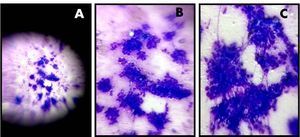
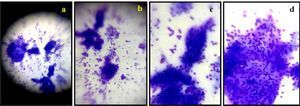
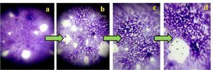
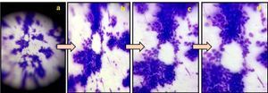
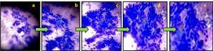
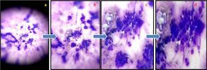
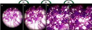
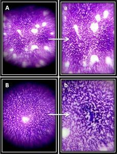
 140x Magnification
 Microorganisms
Ratna Kumari
Learn about the author...
39posts
3comments
1locations
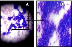
View in Media Gallery
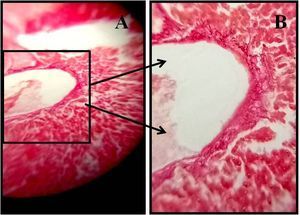
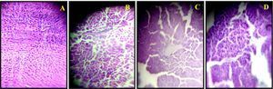
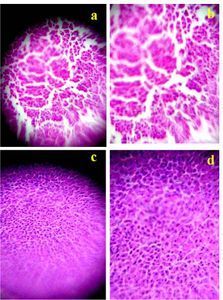
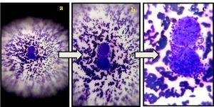
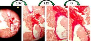
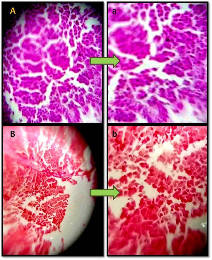
Stained Rat liver section observed under foldscope.
Panel A shows H&E stained DEN treated rat liver section, with disorganized liver architecture, dilated sinusoids, and expanded portal vein. Panel B shows the same section, with Oil red-O staining where the positive red staining is more as lipogenic molecules are more active in this sample. a & b images are zoom out (3X) images of panel A & B respectively.
Panel A shows H&E stained DEN treated rat liver section, with disorganized liver architecture, dilated sinusoids, and expanded portal vein. Panel B shows the same section, with Oil red-O staining where the positive red staining is more as lipogenic molecules are more active in this sample. a & b images are zoom out (3X) images of panel A & B respectively.
Sign in to commentNobody has commented yet... Share your thoughts with the author and start the discussion!
 0 Applause
 0 Comments