Observation of Fine needle aspirate sample of Hepatocellular carcinoma patients under foldscope
 Aug 27, 2019 • 10:45 PM UTC
 Unknown Location
 140x Magnification
 Microorganisms
Ratna Kumari
Learn about the author...
39posts
3comments
1locations
View in Media Gallery
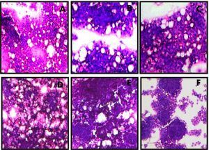
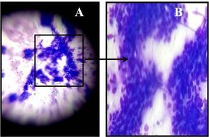
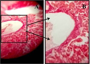
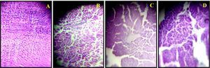
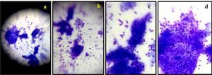
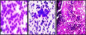
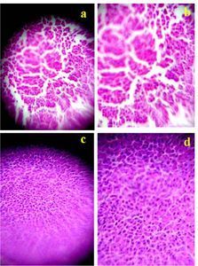
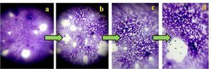
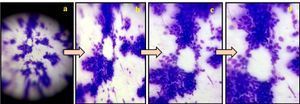
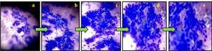
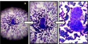
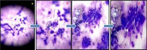
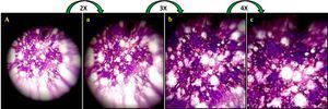
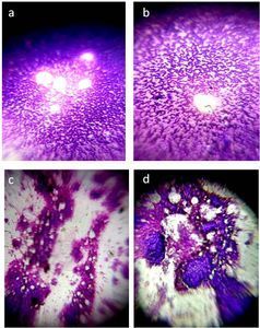
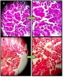
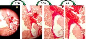
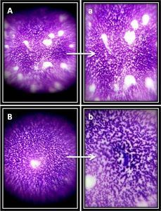
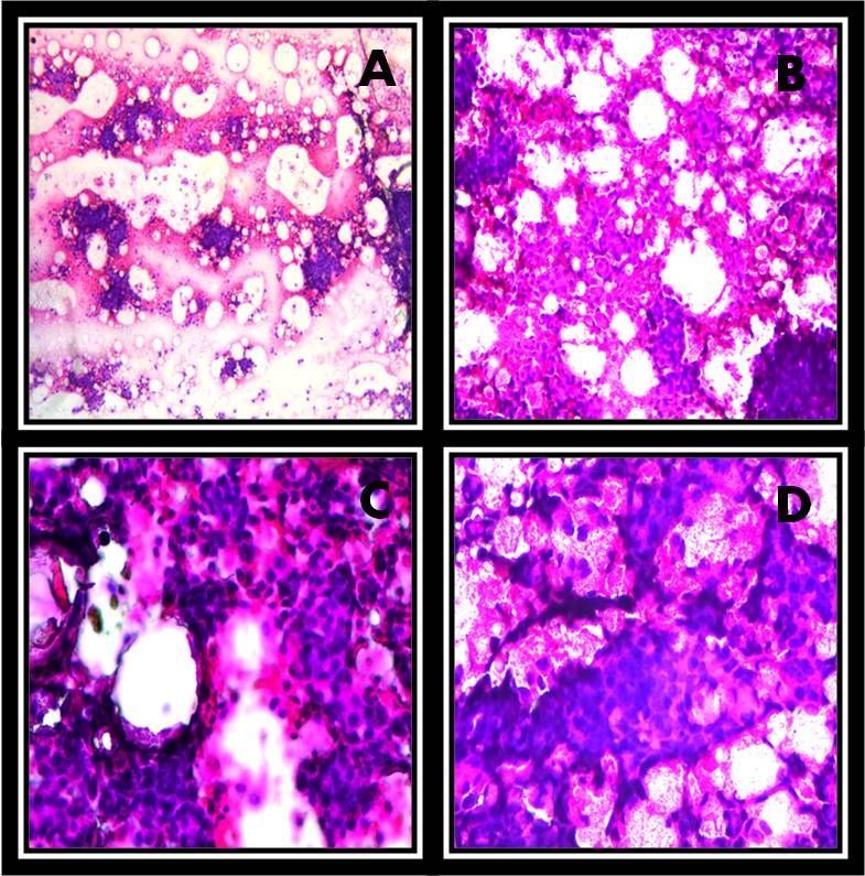
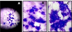
The photomicrographs taken with foldscope demonstrates MGG stained cancer cells in FNAC samples of different stages of different HCC patients. Figure shows distortion of hepatic architecture, Vacuolation, accumulation of lipid droplets. and multilayered cells. Images are the 3X zoom out image taken with OPPO-F7 android cell phone.
Sign in to commentNobody has commented yet... Share your thoughts with the author and start the discussion!
 0 Applause
 0 Comments