Detecting Hepatitis B virus associated cellular changes in needle aspirate samples of HBV linked Hepatocellular carcinoma patients of different stages
 Apr 16, 2019 • 11:26 PM UTC
 Unknown Location
 140x Magnification
 Microorganisms
Ratna Kumari
Learn about the author...
39posts
3comments
1locations
View in Media Gallery
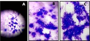
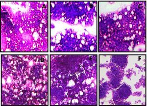
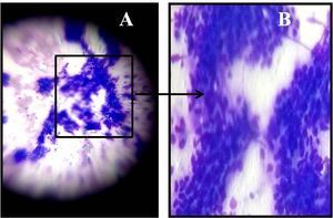
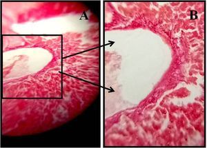
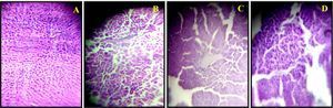
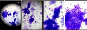
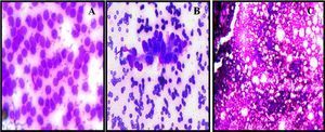
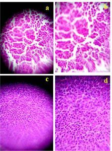
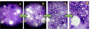
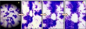
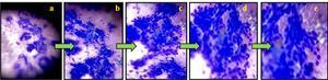
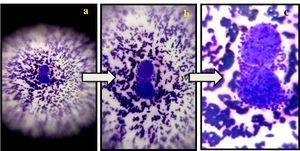
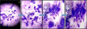
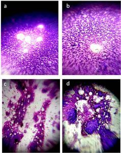
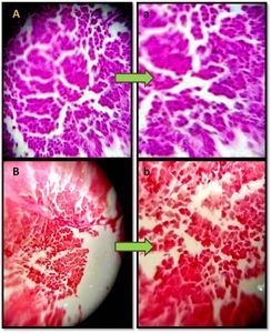
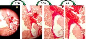
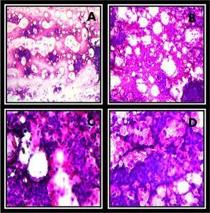
Hematoxylin and Eosin stained sections from Fine Needle Aspirates (FNAC) samples of HCC patients
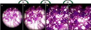
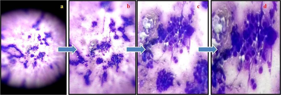
The photomicrgraphs taken with foldscope demonstrates higher stages of HCC patients . Figure a represents cancer cells in FNAC samples of a HCC stages patients which shows cellular clusters of small cells along with widened trabecular, multilayered cells and disorganized architecture. b, c and d are zoom out (b-2X, c-3X and d-4X) images of Fig a.
The photomicrgraphs taken with foldscope demonstrates higher stages of HCC patients . Figure a represents cancer cells in FNAC samples of a HCC stages patients which shows cellular clusters of small cells along with widened trabecular, multilayered cells and disorganized architecture. b, c and d are zoom out (b-2X, c-3X and d-4X) images of Fig a.
Sign in to commentNobody has commented yet... Share your thoughts with the author and start the discussion!
 0 Applause
 0 Comments