Onion Cells
 Nov 27, 2020 • 2:46 AM UTC
 Unknown Location
 140x Magnification
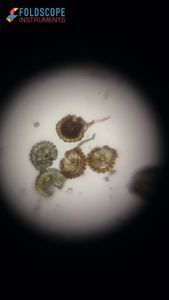
 Microorganisms
Diya A
Learn about the author...
13posts
9comments
1locations
View in Media Gallery
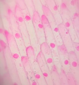
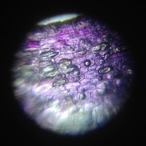
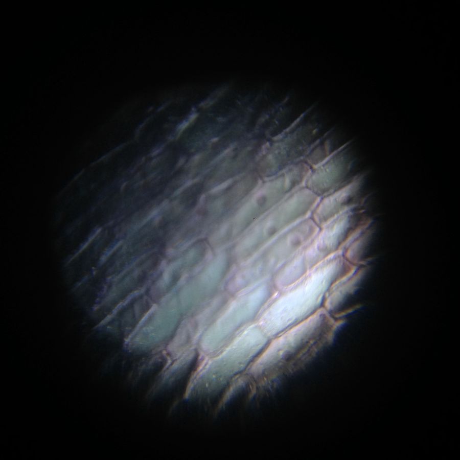
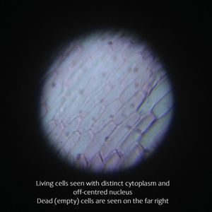
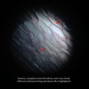
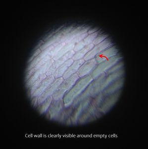
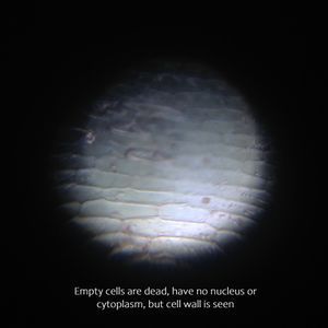
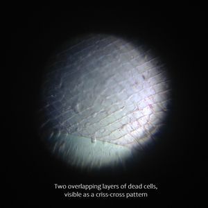
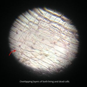
An onion peel is typically one of the first samples viewed when introduced to school-level microscopy. Here are images of an onion skin under my Foldscope. The cells are seen very distinctly and have a three-dimensional appearance.
CookerBird
Sign in to commentNobody has commented yet... Share your thoughts with the author and start the discussion!

 0 Applause
 0 Comments