Bee Wing Sample
 Jun 25, 2026 • 2:33 PM UTC
 India
India 140x Magnification
 Insects and Arachnids
Vismay Anup
Currently studying in the Modern School, ECNCR-Delhi, Vismay Anup is an amateur observer and an aspiring international relations consultant. He loves learning about and studying all sorts of organisms- whether it be microbial or not. Though starting slowly, he is excited to observe as much as possible and learn from other's findings as well. If given free rein, Vismay would be found in one of 6 places: His room, the library, the bookshop, the airport, the zoo and the nearest bio lab.
7posts
3comments
0locations

View in Media Gallery
Hi guys,
This is a bit sudden, but I really felt like sharing these observations with you. Today, I managed to obtain an already prepared sample of a bee wing on a slide and wondered what it would look like on my Foldscope 2.0. So, I pulled it out and observed the wing under both 50x and 140x lenses.
50x
Area Around Tegula
I could observe the membrane of the upper wing, some veins and the tegula itself. As it was 50x, I couldn't observe specific details, but the tegula looked like two small bones creating a rounded shape, probably to fit in the mesonotum . I could also observe hamuli on the membrane.

View in Media Gallery
Area At The Back (Membrane)
Between the membrane, veins ran through the wing to form cell-like patches of membrane. It really reminded me of the onion cells I observed before. Some of the veins were much thicker than others, and sometimes, veins converged to form bigger ones. Hamuli were still observable, though I wasn't too sure if my observations were accurate. Thus, I decided to further observe the wing at 140x.
Between the membrane, veins ran through the wing to form cell-like patches of membrane. It really reminded me of the onion cells I observed before. Some of the veins were much thicker than others, and sometimes, veins converged to form bigger ones. Hamuli were still observable, though I wasn't too sure if my observations were accurate. Thus, I decided to further observe the wing at 140x.

View in Media Gallery
140x
Area Around Tegula
I could observe the axillary sclerites near converging to form the Tegula . I could separately observe the first axillary sclerite and the humeral sclerite. The sclerites were a pale yellow colour, which got deeper as they approached each other, darkest at the base. It kind of reminded me of minuscule human bones.

View in Media Gallery
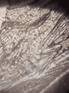
Area At The Back (Membrane)
The first thing that stood out to me was how clear the hamuli were. There were so many of them, looking like hair or small hooks. Previously, I didn't know that they existed, and so, I went online and found out that hamuli are small are tiny, specialised hooks found on the hind wings of bees (and other members of the insect order Hymenoptera ). Their primary purpose is to physically lock the front and back wings together during flight. I was very proud of how clear the picture showed these structures. The veins on the wing were also visible, with a little staining on them (some even leaked out onto the membrane.
The first thing that stood out to me was how clear the hamuli were. There were so many of them, looking like hair or small hooks. Previously, I didn't know that they existed, and so, I went online and found out that hamuli are small are tiny, specialised hooks found on the hind wings of bees (and other members of the insect order Hymenoptera ). Their primary purpose is to physically lock the front and back wings together during flight. I was very proud of how clear the picture showed these structures. The veins on the wing were also visible, with a little staining on them (some even leaked out onto the membrane.

View in Media Gallery
This experience was quite fun and intriguing, as it was my first time observing a part of an animal (insect, to be more specific). I really enjoyed it!
Thanks for reading!
Vismay
Thanks for reading!
Vismay
Sign in to commentNobody has commented yet... Share your thoughts with the author and start the discussion!
_300x300.jpeg)
 0 Applause
 0 Comments