#LG-AUP 2026 Week-3 Day Three (Observations on 3 June 2026) Part 2 - Pollen Grains
 Jun 05, 2026 • 9:29 AM UTC
 India
India 340x Magnification
 Plants
Vismay Anup
Currently studying in the Modern School, ECNCR-Delhi, Vismay Anup is an amateur observer and an aspiring international relations consultant. He loves learning about and studying all sorts of organisms- whether it be microbial or not. Though starting slowly, he is excited to observe as much as possible and learn from other's findings as well. If given free rein, Vismay would be found in one of 6 places: His room, the library, the bookshop, the airport, the zoo and the nearest bio lab.
7posts
3comments
0locations
_900x900.jpeg)
View in Media Gallery
Hello Readers,
Continuing from where I left off in my previous blog, I will be describing my observations made on my Foldscope 2.0 during the latter half of day 3 of the third batch of Exploring the Microcosm course at the LGP at Ashoka University.
In this part of today's workshop, we observed different types of pollen grains and discussed their differences. Our instructor also showed us a little of dark field microscopy. I personally observed the pollen grains of sunflowers and periwinkles, and I will be sharing my observations of the same.
Sunflower Pollen Grain
50x
Even at such a small magnification, individual pollen grains could be observed
All of them were yellowish in colour
They were mostly round and spherical in shape
_900x900.jpeg)
View in Media Gallery
140x
Due to the increase in magnification, I was able to observe a small cluster of about 7 pollen grains
On each pollen grain, multiple hairy structures or hooks were visible. Those are to attach the grains to pollinators, so that pollination can occur.
A yellow tint remained on all grains, and each was structurally almost identical to a sphere.
%20(1)_900x900.jpeg)
View in Media Gallery
340x
Grains were observed using dark-field microscopy, which allowed us to observe individual grains at higher resolution and in greater detail. It also gave the grains a 3D look, making our observations more accurate and astute.
According to Wikipedia, Dark-field microscopy is an illumination technique that enhances the contrast of unstained, transparent samples by illuminating them with obliquely angled light. This causes the specimen to appear brightly lit against a dark, nearly black background. It is highly prized for viewing live, moving microorganisms without altering or killing them.
I feel that Dark-field microscopy really helped us in observing the grains, as without it, the images were quite blurry, but with it, we got such detailed and beautiful images of the pollen grains.
The hook-like hairs were visible, and we noted that there were many covering almost the entire surface of the grain.
The pollen grains became neon in colour, due to the microscopy involved.
Other observations related to the morphology remained the same as previous observations.
%20(1)_900x900.jpeg)
View in Media Gallery
Periwinkle Pollen Grain
50x
At this magnification, individual grains were not visible. Only huge masses/blobs of grains could be observed, which gave it a certain semi-liquid feel.
These groups contained a lot of grains, sometimes overlapping one another.
But still, it was observed that the grains were almost fully transparent with no pigmentation.

View in Media Gallery
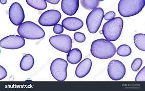
140x
Under this magnification, individual grains were visible but still grouped (in smaller groups of around 9-12 each).
The grains appeared to be ovular-shaped structurally, looking quite like black-eyed beans or lobiya , with certain horizontal markings in the middle of each cell.
One or two cells were also destroyed/flattened in the process of preparing the sample, and so appear as semi-liquid mush.
These grains appear to have a very thick boundary, protecting each grain of pollen.
_900x900.jpeg)
View in Media Gallery
340x
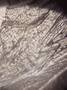
Observed the sample with normal light as well as through dark-field microscopy.
When observing with normal light, the morphology of the pollen was observed.
These grains seem to have a rounded rectangular structure with a line through it.
Certain unidentifiable structures were also observed in each grain.
When observing with dark-field microscopy, we were able to observe the grains as 3-dimensional, with more morphological details.
The pollen grains were transleucent on the barrier, with a darker centre. But, it didn't glow neon like sunflower pollen grains.
_300x300.jpeg)
_300x300.jpeg)
Differences Observed between Sunflower and Periwinkle Pollen Grains
1. The sunflower pollen was spherical in shape, while the periwinkle was more rectangular and bean-shaped.
2. The sunflower pollen grains were much bigger than the periwinkle, as with 50x only, I could observe individual sunflower pollen grains, while for periwinkle, it looked like mush (140x was needed to observe individual grains). Thus, it showed the size difference between the two types of pollen.
3. The sunflower pollen grains had visible hooks extending out of the structure, which were not observable for periwinkle pollen.
4. During dark-field microscopy, we observed that the sunflower pollen grains glowed neon yellow, while the periwinkle grains were dark blue.
5. Sunflower pollen grains had a certain yellow pigment in them, while the pollen of the periwinkle was almost fully transparent.
Challenges Faced
1. Obtaining samples of grains was challenging. Before sunflower, we tried to gain samples of hibiscus, but the specific specimen of hibiscus didn't have any pollen grains which we could obtain, thus we directly moved to sunflower.
2. Spotting cells in 340x, especially for periwinkle, was quite challenging, as we had to change the focus and adjust it multiple times to get a good observation.
3. Trying out dark-field microscopy for the first time was fun, but keeping the foldscope in its place during the process was a little hard.
The day was filled with fun observations as we reached the middle of the week. Though I became a little sad thinking about how little time we had till the end, I resolved to make the most of it and learn as much, so I can perform such observations in my daily life.
Regards,
Vismay
Sign in to commentNobody has commented yet... Share your thoughts with the author and start the discussion!

 0 Applause
 0 Comments