LGP 26- B2 (Day 1) : Fern Rhizome
 May 26, 2026 • 5:00 PM UTC
 India
India 340x Magnification
 Microorganisms
Kavya Poddar
Learn about the author...
7posts
0comments
0locations
View in Media Gallery
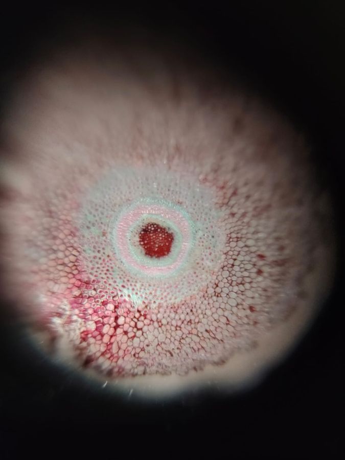
We, at Lodha Genius Programme at Ashoka University, Sonipat, attempted to look at the molecular structure of fern rhizome using the foldscope, at 50x, 140x, and 340x. The rhizome sample was stained and we could easily see the differently coloured cells. We saw it at different magnifications, and in each one we could see more closely inside each cell, and what it looked like. There were multiple rings of cells stained red, blue, and pink. It was my first time using the foldscope and I had some trouble with figuring out how to operate it and how to use the fine adjustment in order to see the sample more clearly.

View in Media Gallery
This is how the rhizome looked like at 50x magnification.

View in Media Gallery
This is how it looked like at 140x.

View in Media Gallery
This is how it looked like at 340x.
This experience was wonderful and it was exhilarating to see the things we see everyday up close and how they're made up of such tiny structures.
This experience was wonderful and it was exhilarating to see the things we see everyday up close and how they're made up of such tiny structures.
Sign in to commentNobody has commented yet... Share your thoughts with the author and start the discussion!
 0 Applause
 0 Comments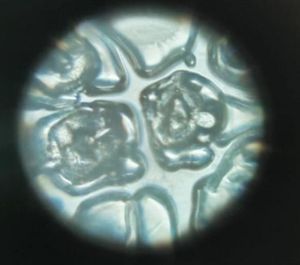
_300x300.jpeg)


