LGP26-B3: Day 1 - Fern Rhizome and Onion Peel
 Jun 05, 2026 • 3:43 PM UTC
 India
India 140x Magnification
 Unknown
Siffat Kaur Kohli
Learn about the author...
12posts
0comments
0locations
View in Media Gallery
In this post I will be continuing the observations for day 1 which include the Fern Rhizome and onion peel under 140X magnification.
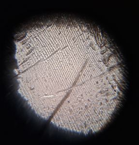
As mentioned earlier, the Fern Rhizome was a permanent slide so the lack of air bubbles and tape made it easier to view under the lens. Here are a few photos of this sample under 140X magnification.
As mentioned earlier, the Fern Rhizome was a permanent slide so the lack of air bubbles and tape made it easier to view under the lens. Here are a few photos of this sample under 140X magnification.


In these images, the cells appear to be larger with less spacing between them. Furthermore, the first picture shows a clear image of the stele core which contains the primary vascular tissue of the Fern Rhizome which is a dark maroon color.
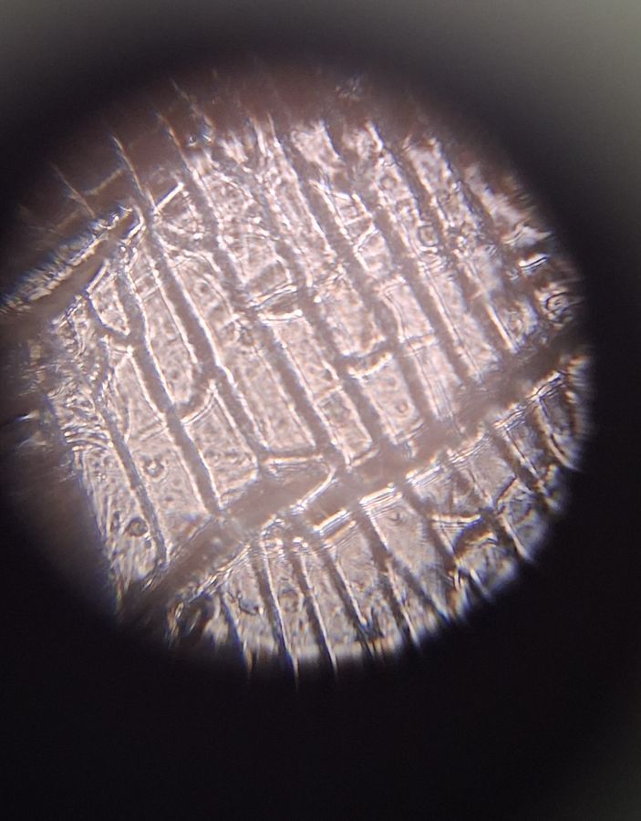
Next, I will talk about the onion peel sample under this magnification. Here are a few photographs.
Next, I will talk about the onion peel sample under this magnification. Here are a few photographs.


I was satisfied with my sample under 140X magnification as it showed a clear view of the cellular structure. The brick-like cellular structure is visible under the layer of tape and the cell wall.
Next, I will be covering the same samples under 340X magnification.
-Siffat Kaur Kohli
Next, I will be covering the same samples under 340X magnification.
-Siffat Kaur Kohli
Sign in to commentNobody has commented yet... Share your thoughts with the author and start the discussion!

 0 Applause
 0 Comments