LGP26-B3: Day 3 - Tomato Peel, Flesh, and Pollen
 Jun 06, 2026 • 6:26 PM UTC
 India
India 340x Magnification
 Unknown
Siffat Kaur Kohli
Learn about the author...
12posts
0comments
0locations

View in Media Gallery
This is my final post for these three samples, the tomato peel, tomato flesh, and pollen.
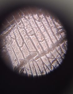
Here are my photographs for the tomato peel.
Here are my photographs for the tomato peel.


The cells, cellular shape, and structure are clearly visible in these photographs. These photos were taken facing a light, hence the light scattering gave it a slightly greenish tint. We can see that the cells are tightly packed and and polygonal.
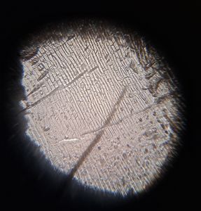
Next, the tomato flesh sample.
Next, the tomato flesh sample.

View in Media Gallery
We can see the parenchyma cells and the sample seems to be semi transparent.
Finally, the pollen samples.
The sunflower pollen was circular, bright yellow, and had thick black spikes around it.
The frangipani and periwinkle sample showed clear lines within the semi-transparent, coffee bean cells.
In my next post, I will be covering a leaf sample.
-Siffat Kaur Kohli
Finally, the pollen samples.
The sunflower pollen was circular, bright yellow, and had thick black spikes around it.
The frangipani and periwinkle sample showed clear lines within the semi-transparent, coffee bean cells.
In my next post, I will be covering a leaf sample.
-Siffat Kaur Kohli
Sign in to commentNobody has commented yet... Share your thoughts with the author and start the discussion!

 0 Applause
 0 Comments