LGP26-B3: Day 3 - Tomato Peel, Flesh, and Pollen
 Jun 06, 2026 • 5:47 PM UTC
 India
India 50x Magnification
 Unknown
Siffat Kaur Kohli
Learn about the author...
12posts
0comments
0locations

View in Media Gallery
In this post, I will be covering the samples prepared on day 3 of the workshop, tomato peel and tomato flesh.
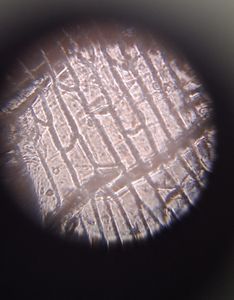
First, we got a sample of the tomato peel by peeling off a thin sample using our nails and then taping them to the slide.
First, we got a sample of the tomato peel by peeling off a thin sample using our nails and then taping them to the slide.
View in Media Gallery
The cells in this image are compact and have a reddish tint to them. Although it is not clearly visible in this photo, the cells have a polygonal structure. These are epidermal cells with thick walls.
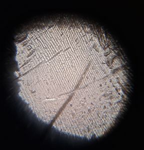
Next, the tomato flesh. I took a small piece of tomato flesh, grinded it, took away the excess pulp, and taped it onto a slide.
Next, the tomato flesh. I took a small piece of tomato flesh, grinded it, took away the excess pulp, and taped it onto a slide.

View in Media Gallery
There are parenchyma cells which store water. They are large, circular, and they are also thin-walled. We can also see the nuclei inside the parenchyma cells.
Finally, we saw pollen samples from three different flowers; Sunflower, Periwinkle, and Frangipani.
My pictures for these samples were rather unclear, hence I will be typing out my observations instead.
The sunflower pollen was small, circular, yellow, and seemed to have faint spikes around it.
The periwinkle and frangipani samples looked nearly identical. They were both colorless, elliptical, and resembled coffee beans.
In my next two posts, I will be covering the same samples under 140X and 340X magnification.
-Siffat Kaur Kohli
Sign in to commentNobody has commented yet... Share your thoughts with the author and start the discussion!

 0 Applause
 0 Comments