LGP26-B3: Day 1 - Fern Rhizome and Onion Peel
 Jun 02, 2026 • 10:26 AM UTC
 India
India 50x Magnification
 Unknown
Siffat Kaur Kohli
Learn about the author...
12posts
0comments
0locations
View in Media Gallery
In this workshop, we observed a sample of Fern Rhizome as well as an onion peel using our foldscopes. These samples were viewed through three lenses: 50X, 140X, and 340X. In this blog post, I will only be covering magnification 50X.
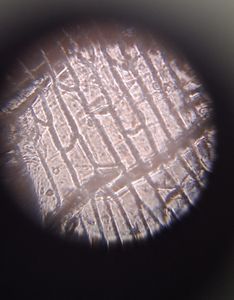
The Fern Rhizome was a permanent and pre-stained slide, so we did not have to prepare it before inserting it towards the lens. A Fern Rhizome is the main stem of a fern and in my opinion, has an interesting tissue structure. Once the lens was secured, I held it up to the light and attempted to take a few photographs from different angles. Here are the pictures:
The Fern Rhizome was a permanent and pre-stained slide, so we did not have to prepare it before inserting it towards the lens. A Fern Rhizome is the main stem of a fern and in my opinion, has an interesting tissue structure. Once the lens was secured, I held it up to the light and attempted to take a few photographs from different angles. Here are the pictures:


Taking the photos posed a challenge as it is difficult to hold the foldscope still, however I have inserted several examples from different angles to ensure that the reader gets some clarity as to how this sample looks under this magnification. The epidermis has a blue tint and consists of what seem to be pavement cells. The cells are shaped like irregular polygons, especially irregular hexagons. The colors are blue, pink, purple, and red.
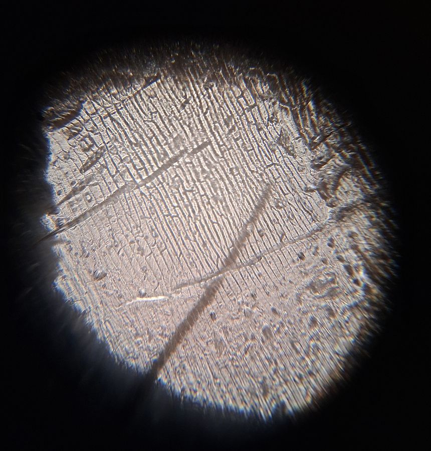
Next, we made our own onion peel samples. We peeled off the outer layer of an onion and stuck it with tape on a slide. Then, I proceeded to take pictures and note down my observations. I will only be inserting one image as it gives a clear idea about how onion cells look under this magnification.
Next, we made our own onion peel samples. We peeled off the outer layer of an onion and stuck it with tape on a slide. Then, I proceeded to take pictures and note down my observations. I will only be inserting one image as it gives a clear idea about how onion cells look under this magnification.

View in Media Gallery
This picture was taken while directly facing the light so the nuclei and cell walls are visible. The nuclei resemble blobs on top of the brick-like cell structure while the cell walls look like horizontal squiggly lines which disrupt the black and white cell pattern.
These were my observations of these two samples under 50X magnification. In my next two posts, I will be covering the same samples under 140X and 340X magnification.
-Siffat Kaur Kohli
These were my observations of these two samples under 50X magnification. In my next two posts, I will be covering the same samples under 140X and 340X magnification.
-Siffat Kaur Kohli
Sign in to commentNobody has commented yet... Share your thoughts with the author and start the discussion!

 0 Applause
 0 Comments