LGP26-B3: Day 3 - Tomato Peel, Flesh, and Pollen
 Jun 06, 2026 • 6:10 PM UTC
 India
India 140x Magnification
 Unknown
Siffat Kaur Kohli
Learn about the author...
12posts
0comments
0locations

View in Media Gallery
I will be discussing the tomato peel, tomato flesh, and pollen samples covered on day 3 under 140X magnification.
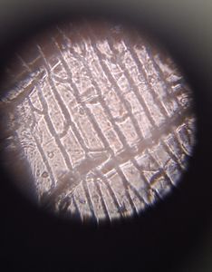
Here are my photographs for the tomato peel.
Here are my photographs for the tomato peel.


These pictures are much clearer and the polygonal structure of the cells is more visible and discernible. We can also see some air bubbles from the tape.
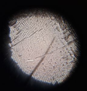
Next, here are my images for the tomato flesh.
Next, here are my images for the tomato flesh.


The parenchyma cells are enlarged. It is also important to note that there were some air bubbles from the tape in my sample, so it might be slightly confusing however I have gotten images of the cellular structure.
Finally, the pollen samples.
The sunflower pollen had more distinct spikes under this magnification.
The frangipani and periwinkle pollen seemed to have slight lines inside their coffee bean shape.
In my next post, I will finish these samples and move on to my final leaf sample.
-Siffat Kaur Kohli
Finally, the pollen samples.
The sunflower pollen had more distinct spikes under this magnification.
The frangipani and periwinkle pollen seemed to have slight lines inside their coffee bean shape.
In my next post, I will finish these samples and move on to my final leaf sample.
-Siffat Kaur Kohli
Sign in to commentNobody has commented yet... Share your thoughts with the author and start the discussion!

 0 Applause
 0 Comments