LGP26-B3: Day 2 - Potato Peel and Potato Flesh
 Jun 06, 2026 • 4:18 PM UTC
 India
India 50x Magnification
 Unknown
Siffat Kaur Kohli
Learn about the author...
12posts
0comments
0locations

View in Media Gallery
In this workshop, we observed potato peel and potato flesh samples under the foldscope. We viewed them through three lenses, 50X, 140X, and 340X. In this post, I will only be covering 50X magnification.
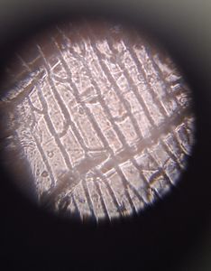
To prepare the potato peel sample, we peeled a thin layer off a potato and stuck it onto a slide with tape. Here is an image.
To prepare the potato peel sample, we peeled a thin layer off a potato and stuck it onto a slide with tape. Here is an image.
View in Media Gallery
The potato peel (periderm) had compact cells which were easily visible under this magnification. There was a distinct dark brown pigmentation. The phellem (or cork cells) are dead cells which act as a protective layer.
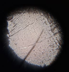
Next, we prepared the potato flesh sample, which proved to be more difficult. We cut out a small piece of the potato flesh, grinded it, and taped the transparent remains on the slide. Here are the images.
Next, we prepared the potato flesh sample, which proved to be more difficult. We cut out a small piece of the potato flesh, grinded it, and taped the transparent remains on the slide. Here are the images.


In the potato flesh, we can see compact parenchyma cells which contain starch grains. The starch grains resemble ovals and are semi-transparent.
In my next two posts, I will be covering the same samples under 140X and 340X magnification.
-Siffat Kaur Kohli
In my next two posts, I will be covering the same samples under 140X and 340X magnification.
-Siffat Kaur Kohli
Sign in to commentNobody has commented yet... Share your thoughts with the author and start the discussion!

 0 Applause
 0 Comments