LGP26-B3: Day 1 - Fern Rhizome and Onion Peel
 Jun 05, 2026 • 4:06 PM UTC
 India
India 140x Magnification
 Unknown
Siffat Kaur Kohli
Learn about the author...
12posts
0comments
0locations
View in Media Gallery
This is my final post for Day 1 of the microcosm workshop. In this blog, I will be covering the Fern Rhizome and onion peel samples under 340X magnification.
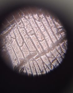
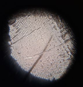
The Fern Rhizome, the main stem of a fern, was clearly visible under this lens. We got a closer look at the zoomed-in cellular structure and I have taken several pictures.
The Fern Rhizome, the main stem of a fern, was clearly visible under this lens. We got a closer look at the zoomed-in cellular structure and I have taken several pictures.


The shading in the pavement cells located in the epidermis is clearly visible and all previous observations covered are visible here in more detail.
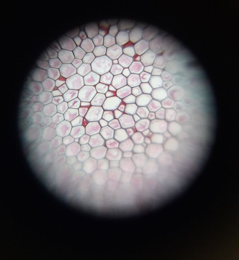
Finally, we looked at the onion peel under 340X magnification. Here are the photographs I clicked.
Finally, we looked at the onion peel under 340X magnification. Here are the photographs I clicked.


The brick-like cellular structure is enlarged and clearly visible. In this sample as well, all previously stated observations in my previous two posts were visible however these are focused images. Next, I will be covering the potato peel and flesh.
-Siffat Kaur Kohli
-Siffat Kaur Kohli
Sign in to commentNobody has commented yet... Share your thoughts with the author and start the discussion!

 0 Applause
 0 Comments