Culturing your favorite ciliate: Paramecium busaria
 Apr 11, 2019 • 9:10 PM UTC
 Unknown Location
 140x Magnification
 Microorganisms
Laks Iyer
Human observer of life. https://sukshmadarshin.wordpress.com
97posts
1255comments
5locations
View in Media Gallery
It happens to all of us collectors. You find an interesting rock and you want to keep it in your pocket for further study. Many kids have pencil boxes and pockets full of bugs. In my case, I dream to have a large collection of ciliates, although I have had more failures than success in this process, and it is frustrating at times to see your efforts go waste. Yet, the ones that do grow bring tremendous joy. The most popular and readily available medium for this exercise is the wheat medium. Very simply, in about 250 ml of “Spring water” (I just get a big can of bottled spring water) drop 4-8 boiled wheat berries to give wheat medium. I always keep a stock of sterile bottled water and a bottle of cooked wheat berries. I basically pressure cook the two when no one is at home and it lasts for months at times.
Once I spot an interesting ciliate, I will almost instinctively throw it into a glass petriplate or a tube with this medium and observe the plate for up to a month. Most of the times one faces disappointment, but “when the stars align” one might get an explosive growth of the ciliate of choice. Recently, I put a wheat berry in a pond water sample and kept it under 12 h light and 12 h darkness. In about 2 weeks, there were green ciliates growing vigorously as seen in the video below. On consulting books and experts, I realized it was Paramecium bursaria.
Once I spot an interesting ciliate, I will almost instinctively throw it into a glass petriplate or a tube with this medium and observe the plate for up to a month. Most of the times one faces disappointment, but “when the stars align” one might get an explosive growth of the ciliate of choice. Recently, I put a wheat berry in a pond water sample and kept it under 12 h light and 12 h darkness. In about 2 weeks, there were green ciliates growing vigorously as seen in the video below. On consulting books and experts, I realized it was Paramecium bursaria.
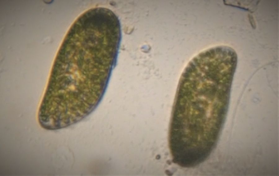
Paramecium bursaria growing in a petridish. Note the large number of ciliates in one field of view. Under the foldscope Paramecium bursaria is delightful. Here I have mixed it with Paramecium multimicronucleatum for comparison. What strikes me is that these are two members of the same genus, yet the gross features are so different.
Note how the ciliates scan each other. At times antagonistic interactions result in release of toxic barbs called trichocysts. What might be the molecular basis for this? The third and really small ciliates is likely to be Cyclidium Note how ciliates scan each other intimately. Manu calls this the ciliate handshake. See https://microcosmos.foldscope.com/?p=30093
In the following video we see the flow field of the vortex created by Paramecium .
In the following video we see the flow field of the vortex created by Paramecium .
A closer look at the ciliate expounds a principle of biological conflict seen across many scales of life in biology, perhaps best summarized for this post as Capture . The Paramecium seen here captured an alga of the Zoochlorella genus, which is a eukaryotic alga whose ancestor about 1.6 billion years ago captured a cyanobacterium to give rise to the plant lineage, which was captured under a foldscope and whose image was captured in my eye and on the phone video. Note that each alga is enclosed in a separate vacuole.
Finally, there is a virus that infects the Zoochlorella alga, in Paramecium bursaria called Paramecium busraria chlorella virus. Many years ago, my colleagues and I realized while studying its genome that this virus and many other giant viruses have a common origin and we called these the Nucleo-cytoplasmic Large DNA viruses. This study changed the way proteins and genomes of these large viruses were studied. In a sense getting Paramecium bursaria from my local pond was a very satisfying moment for me.
P,S. I have a dense growth of this ciliate. If you have any experiments you think I could do in a home lab, I will be happy to try it.
P,S. I have a dense growth of this ciliate. If you have any experiments you think I could do in a home lab, I will be happy to try it.
Sign in to commentNobody has commented yet... Share your thoughts with the author and start the discussion!

 0 Applause
 0 Comments
_300x300.jpeg)