Experiments with Blood
 Jan 14, 2015 • 2:35 PM UTC
 Unknown Location
 140x Magnification
 Unknown
Laks Iyer
Human observer of life. https://sukshmadarshin.wordpress.com
97posts
1255comments
5locations
View in Media Gallery
I was gifted my first bulb microscope at 8. It was a simple microscope but gave fascinating views of the unknown. One of my companions in crime pushed a pin in his finger and dropped some blood onto the only slide that came with the microscope. We fought and took turns to see if we could spot something. All we could see was a red mess. Viewing blood cells under a microscope was much harder than I thought. Indeed I learnt much later that even a small volume of blood (1 microliter) has between 4-6 million Blood cells, of which the red blood cells (RBCs) or erythrocytes are the most abundant. That explained the mess.
Now with the foldscope, it is time to revisit blood. I am going to try a variety of experiments to see which gives me the best resolution and hence this is likely to be a post spanning a few days/weeks.
1) For the first experiment, I punctured my finger with a lancet (always use a sterile one, these are easily available, and you can always get a disposable one from an uncle), put a drop of blood at one end of the slide. With another slide I made a smear than thinned across the slide. For a pictorial example as to how to do it, see http://www.cdc.gov/dpdx/resources/pdf/benchAids/malaria/Malaria_procedures_benchaid.pdf
I let the smear air dry and put a drop pf 95% ethanol that I had a supply of for my spirit lamp (very easy to get from Home depot). After a minute, I poured it off and added 1% Methylene Blue for about 5 minutes and washed it off with tap water. The smear generally sticks well to the slide after the air drying. This process is nowhere close to what the experts use with Romanowsky or Wright stains and so I did not expect to see the various white blood cells, but red blood cells I knew I would see. Here is my first view of Red Blood Cells (erythrocyes). these are snapshots taken that were collated in picasa. The magnification is 140x plus 4x digital zoom from the phone; so around 560x.
View in Media Gallery
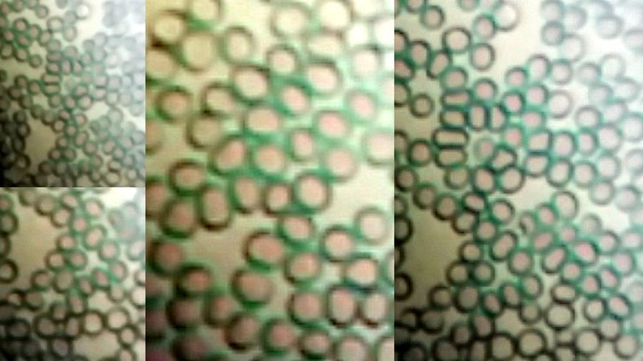
The red color of the red blood cells (erythrocytes) is seen clearly. Human RBCs do not have a nucleus (no excess baggage) and hence the methylene blue stain didnt show any nucleus as it does say with the cheek cells. Most of the slide was filled with RBCs, for after all there are 4-6 million red blood cells per microliter.
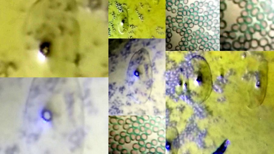
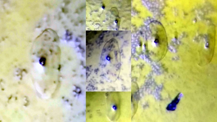
White blood cells were almost nowhere to be seen and I suspect my staining procedure was too simplistic compared to those of Wright and Romanowsky. “ However, there were a few remarkable views for which I had to thoroughly scan the slides. I did find some transparent cells with polymorphic nuclei. These are likely to be the most abundant WBCs, the neutrophils, but their outline suggests an enormous size. If you look carefully in the video and the pictures below, in each case, there is an internal outline/ring surrounding the nuclear material stained blue, which corresponds to the neutrophil’s real size. The outer outline/ring is quite strange. Perhaps the ethanol treatment used as a fixer caused some lysis resulting in some leakage? Dont know. (Recommendations and ideas are always welcome)..”
I realized that the potential WBCs (wrritten in bold and italicized above) was an artifact when I noticed the same bleb in other slides. I did manage to get WBCs though and this is in another post.
View in Media Gallery
The following video summarizes the findings. (To be continued) …
Sign in to commentNobody has commented yet... Share your thoughts with the author and start the discussion!

 0 Applause
 0 Comments
_300x300.jpeg)


